Body regions: what is where
Recognize the main regions of the dog's body.
The interactive dog atlas
Discover the parts of a dog's body and what they do. You can start step by step or go straight to what interests you.
33 body regions, 27 muscles, 39 bones and 33 organs on the same drawing · 13 short lessons · 105 verified questions. Educational material, not a diagnostic tool: for your dog's health, talk to your veterinarian. Version 1.2.0, updated 28.10.2026.
Tap a part of the dog or search for it by name; the explanation appears next to the drawing. The view is the left side: head to the left, the dog's left side facing you.
Results appear below the field; Enter picks the first result.
Tap a part of the dog to find out what it is.
Step by step ·
The interactive explorer could not start in this browser. The drawing and the cards below remain available.
Learning paths
Three lessons to start with: the parts of the body, the joints and the bones. Each takes 3-5 minutes, has a few steps on the drawing and a few questions at the end; mistakes are explained, with no timer and no leaderboard. The lesson opens next to the drawing. Just want to test yourself? Every lesson also has a “Questions only” button.
“Beginner”: questions you answer from the drawing and the lesson, suitable for middle school too. “Advanced”: all the questions, including those with terms and numbers.
Recognize the main regions of the dog's body.
Tell apart the stifle, the hock, the elbow and the carpus (the joint above the front paw).
Recognize the dog's main bones and how they are connected to one another.
Understand how muscles attach to bones (origin, insertion), which ones carry the trunk, which ones push and which ones breathe.
Tell the dog's gaits apart by the sequence in which the limbs touch the ground.
Know what touches the ground in a dog and what does not: pads, toes, dewclaw, carpal pad, claws.
Know where the major organs sit in the dog's body, relative to the ribs and the vertebrae, and why some are drawn with a dashed outline.
Know the categories of teeth, their number in the two dentitions and what a normal bite means.
Follow the path of sniffed air from the nostril to the olfactory bulb and understand why the dog's nose is built for smell.
Recognize what the tail, the ears, the posture and the coat communicate, with the evidence behind each signal.
Measure height and length correctly, tell sexual maturity from the end of growth, and understand what the coat is made of.
Recognize the regions of the head, then see, layer by layer, what sits under them: the bones of the face, the muscles that move the lips and the nose, the glands and the brain.
Follow the scent signal from the olfactory bulb to the areas that identify it, give it value, remember it and decide, and know what has been measured in the dog and what is only a hypothesis.
Challenge of the day · 29.10.2026
Pick the structure from the list below or .
Pelvis (hip bone). The pelvis is formed by the two hip bones, below the croup, between the sacrum and the femur; the head of the femur fits into its socket.
Your progress
0 of 132 structures explored
0 of 13 lessons completed
Remembered only while the page is open. Nothing is saved in the browser until you choose to.
What gets saved if you choose this: the structures explored, the lessons completed with their score, whether you have seen the tour and the level you chose. They stay only in your browser, with no account, no name and nothing sent to us; you can delete them from here at any time.
Topics
Each topic also has a page of its own, with the full text: Exterior, Skeleton, Teeth, Movement, Measurements, Skin and coat, Internal systems, Scent, Behavior, Senses, Muscles, Skull, Brain, Head.
Permanent dentition: 2 × (I 3/3, C 1/1, P 4/4, M 2/3) = 42. Deciduous (milk) dentition: 2 × (i 3/3, c 1/1, p 3/3) = 28.
When the teeth come in
Average ages, with considerable variation; very small breeds tend to be late.
Occlusion
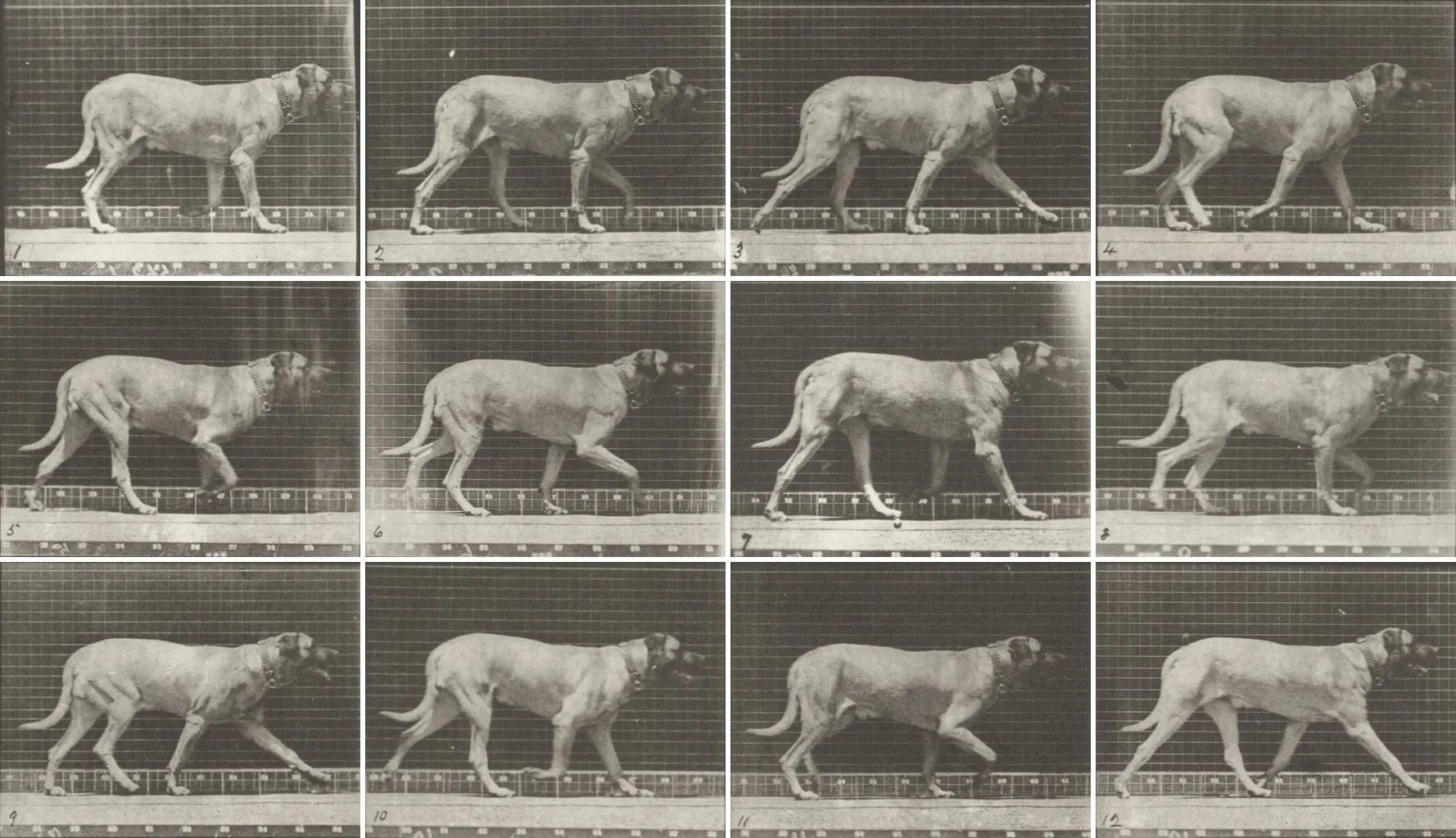
Walk. Four beats: each limb lands in turn, in lateral sequence (a hind limb is followed by the forelimb on the same side). There are alternately two and three limbs on the ground; for each limb the stance lasts longer than the swing.
The stance durations in the strip are a teaching scheme.
Walk. Four beats: each limb lands in turn, in lateral sequence (a hind limb is followed by the forelimb on the same side). There are alternately two and three limbs on the ground; for each limb the stance lasts longer than the swing.
Trot. Two beats: the diagonals alternate (right fore + left hind, then left fore + right hind), with short suspension phases between them. The hind limb of the diagonal touches the ground slightly before the forelimb.
Pace. Two beats with lateral pairs: the two limbs on the same side carry the weight alternately. In dogs it is often seen in tired, overweight or untrained animals.
Canter (slow gallop). Three beats, an asymmetric gait: one hind limb, then a diagonal pair together, then the “leading” forelimb. Suspension appears only at high speed. Often slower than the trot; it shifts easily into the transverse gallop when the diagonal splits.
Transverse gallop. Four beats with a single suspension: the hind pair, then the fore pair, in the same left-right order (for example left hind, right hind, left fore, right fore, with the right forelimb “leading”). The suspension comes after the leading forelimb leaves the ground. It is the horse's gallop; dogs use it too.
Rotary gallop. Four beats with two suspensions: right hind, left hind, extended suspension, left fore, right fore, gathered suspension. It is the dog's fastest gait, but also the most tiring; flexion and extension of the spine lengthen the stride a great deal. A greyhound can reach about 60 km/h.
Pick a measurement: its line appears on the drawing, and the explanation of the landmarks here.
The drawing has no scale: it does not calculate centimeters and does not recommend harness sizes. Full page about measurements.
The same outline — the atlas drawing — scaled to the height at the withers required by each breed's FCI standard (1 cm = 4 units on the ruler) and, where the standard gives a numeric ratio, stretched in length. Pick the breeds and compare them.
| Breed | Standard | Height at the withers | Length : height | Other stated proportions |
|---|---|---|---|---|
| Great Dane | No. 235, 21.08.2024, group 2 | males at least 80 cm (at most 90), females at least 72 cm (at most 84) | almost square, especially in males; females can be a little longer | |
| Greyhound | No. 158, 08.09.2026, group 10 | ideal: males 71-76 cm, females 68-71 cm | the standard gives no ratio; “generous proportions”, long head and neck, deep chest | |
| Bloodhound | No. 84, 13.03.2001, group 6 | ideal 68 cm males, 62 cm females (tolerance 4 cm either way) | body length : height at the withers = 10 : 9 | Depth of chest : height at the withers = 1 : 2; Length of head : length of body = 3 : 7; Length of muzzle : length of head = 1 : 2 |
| German Shepherd Dog | No. 166, 11.08.2010, group 1 | males 60-65 cm, females 55-60 cm | the trunk exceeds the height at the withers by about 10-17 % | Length of head: about 40 % of the height at the withers; Depth of chest: about 45-48 % of the height at the withers |
| Belgian Shepherd Dog | No. 15, 13.03.2001, group 1 | ideal, on average: 62 cm males, 58 cm females (limits: 2 cm under, 4 cm over) | “fits into a square” | The chest reaches down to the level of the elbows; Muzzle: equal to or slightly longer than half the length of the head |
| Golden Retriever | No. 111, 08.09.2026, group 8 | males 56-61 cm, females 51-56 cm | the standard gives no ratio | Skull: broad, without coarseness |
| Siberian Husky | No. 270, 14.11.2022, group 5 | males 53.5-60 cm, females 50.5-56 cm; weight males 20.5-28 kg, females 15.5-23 kg | the body (from the point of the shoulder to the point of the croup) slightly longer than the height at the withers | Distance from nose to stop equal to the distance from stop to occiput; Leg from elbow to ground: slightly more than the distance from elbow to withers |
| Labrador Retriever | No. 122, 08.09.2026, group 8 | ideal: males 56-57 cm, females 54-56 cm | the standard gives no ratio; “strongly built, short-coupled” | Skull: broad, clean-cut, without fleshy cheeks |
| Border Collie | No. 297, 24.06.1987, group 1 | ideal: males 53 cm, females slightly less | body slightly longer than the height at the shoulder | Skull and foreface approximately equal in length |
| Whippet | No. 162, 08.09.2026, group 10 | desirable: males 47-51 cm, females 44-47 cm | the standard gives no ratio | Skull: long and lean, flat on top, tapering toward the muzzle, with a slight stop |
| Beagle | No. 161, 08.09.2026, group 6 | 33-40 cm (desirable minimum and maximum, no difference between the sexes) | the standard gives no length : height ratio | Head divided as equally as possible by the stop; Height to elbow: about half the height at the withers |
| French Bulldog | No. 101, 10.08.2023, group 9 | males 27-35 cm, females 24-32 cm (tolerance 1 cm); weight males 9-14 kg, females 8-13 kg | body length (point of the shoulder - point of the buttock) slightly exceeds the height at the withers; short face | |
| Dachshund (Teckel) | No. 148, 25.05.2021, group 4 | the standard gives no height: size is set by chest girth, measured after 15 months | body length (from the manubrium of the sternum to the point of the buttock) : height at the withers ≈ 1.7-1.8 : 1 | Ground clearance under the body: about one third of the height at the withers |
| Chihuahua | No. 218, 04.09.2019, group 9 | the standard goes by weight, not height: 1-3 kg (ideal 1.5-2.5 kg) | body slightly longer than tall; an almost square body is desired, especially in males | “Apple-shaped” head, well rounded |
| Bulldog | No. 149, 08.09.2026, group 2 | the standard gives no height | the standard gives no ratio | Skull: relatively large in circumference; viewed from the front, high and broad, square |
The FCI standards describe what the breed club requires for the show ring, not anatomy and not health; the real shape of each breed (head, ears, coat, angulation) is not drawn — only the declared size and proportion, on the outline of a male in the middle of the range. Breeds with no height in the standard (Dachshund, Chihuahua, Bulldog) appear only in the table.
Ground-side (palmar) view of the left forepaw, schematic. The dog is digitigrade: it walks on its toes, with the metacarpus raised off the ground; that is why the carpal pad and the dewclaw sit above the ground.
Pick a part of the paw from the drawing or from the buttons.
The metacarpal pad sits under the metacarpophalangeal joints, the digital pads under the last joints of the toes; they are the ground-contact surfaces of a digitigrade animal.
One pad on each of digits II-V, under the joint of the last phalanx. They are made of connective and fatty tissue, which cushions, and of a thick, horny epidermis; the layer that touches the ground has a “honeycomb” structure that does not exist on the side walls of the pad. The pads contain eccrine sweat glands, with at most a minor role in cooling: the dog cools mainly by panting.
A single shared pad, behind the toes, under the metacarpal bones (on the hind paw, the metatarsal pad). It has the same structure as the digital pads: cushioning fatty tissue under a thick epidermis.
The pad on the back of the forelimb, above the accessory carpal bone; veterinary dictionaries also call it the “stopper pad”. It exists only on the forelimbs and normally bears no weight: the dog walks on its toes, with the metacarpus raised. The claim that it touches the ground at the gallop or when braking has no verified source.
The rudimentary digit I, on the inner side of the forepaw, above the weight-bearing toes; it takes no part in support and has its own muscle (the long abductor of digit I). In some puppies it is removed at birth. On the hind limbs it appears only occasionally; in a few breeds the standard requires hind dewclaws, sometimes double. A dog without a dewclaw has digits 2-5.
The four weight-bearing toes, each with three phalanges, a pad and a claw. The dog stands on them, like a runner on tiptoe: the metacarpus stays raised off the ground. When running, each limb takes ground reaction forces of two to three times body weight.
The claw is a horny sheath over the ungual process, the conical, flattened end of the last phalanx. The root of the claw sits under a crescent-shaped bony ridge, the ungual crest, from which it grows.
From the nose leather to the areas of the brain that decide, on two drawings: the section through the head and the brain in lateral view. The atlas's Mantrailing extension: what happens in the dog's nose and head when it sniffs a trail.
1 / 19 · in the nose
1. The nose leather: cold, wet and heat-sensitive
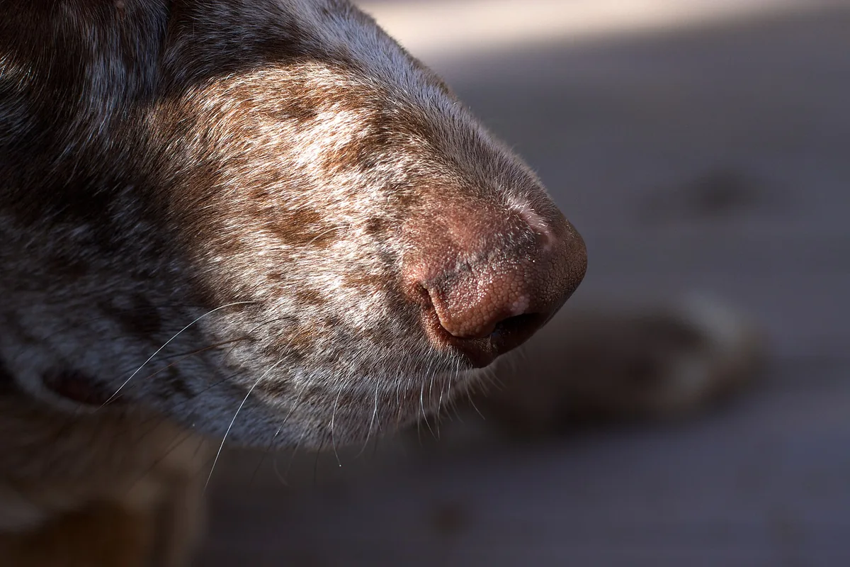
The nose leather, the planum nasale, is the bare, pigmented skin at the tip of the nose, wet and cold in a healthy dog, with a pattern of grooves unique to each dog; it is richly innervated by the trigeminal nerve. Three trained dogs chose, from 1.6 m, a surface only 11–13 °C warmer than the air, and in 13 dogs scanned with fMRI the warm stimulus activated the left somatosensory association cortex. How exactly the nose leather senses heat is not yet known; its moisture does not “catch” scent molecules and, according to the authors, does not cool the body either.
Tap any structure on the drawing: those with a step of their own jump to their step, the others show their card.
The nose leather, the planum nasale, is the bare, pigmented skin at the tip of the nose, wet and cold in a healthy dog, with a pattern of grooves unique to each dog; it is richly innervated by the trigeminal nerve. Three trained dogs chose, from 1.6 m, a surface only 11–13 °C warmer than the air, and in 13 dogs scanned with fMRI the warm stimulus activated the left somatosensory association cortex. How exactly the nose leather senses heat is not yet known; its moisture does not “catch” scent molecules and, according to the authors, does not cool the body either.
When it searches, the dog sniffs in bursts of 4–7 per second; the frequency does not depend on body size, the airflow does. Four German shepherds on a trail sniffed about 6 times per second in all phases of the search. Sniffing sends the air in a laminar, one-way flow toward the dorsal meatus and the ethmoturbinates; that is why it is more efficient for smelling than an ordinary inhalation.
At the peak of inhalation each nostril “pulls” air from an area of about 1 cm in front of it, smaller than the distance between the nostrils: the nostrils take separate samples, from the left and from the right, which helps locate the source. The wing of the nostril, the mobile upper edge of the comma-shaped nostril, is moved by the levator nasolabialis. The dog usually starts with the right nostril; with a new, non-threatening scent it switches to the left when it meets it again, and with adrenaline or the scent of a veterinarian's sweat it stays on the right.
Exhaled air leaves the nasopharynx through the respiratory region and exits at the nostril; because of the internal shape of the nasal vestibule, it comes out as a jet directed downward and to the side, with two vortices, not straight back over the trail. The jet stirs up scents from the ground, which can then be inhaled, without scattering the sample. On exhalation no air enters or leaves the olfactory recess. The phrase “lateral slits of the nostrils” does not appear in the sources we read.
In the vestibule the inhaled air is turbulent and well mixed; then the flow splits. A single path, the dorsal meatus, carries odorant-laden air to the olfactory part of the nose, bypassing the respiratory passages: in Craven's (2010) computer model, about 12–13 % of the inhaled air reaches the chemosensory region. The rest is directed by the respiratory passages toward the nasopharynx. Olfactory and respiratory airflow are separate phenomena.
The ventral nasal concha, the maxilloturbinate, attaches to the maxilla and fills the front part of the cavity; in carnivores it is of the branched type, short and very complex, with coiled lamellae, and the ventral meatus is very narrow alongside it. Above, the dorsal concha, the longest, borders the dorsal meatus. The conchae humidify, warm and filter the air going to the lungs; there are only a few olfactory neurons here. This path leads to the pharynx, not to the brain.
The dog does not cool itself by sweating but by panting, mouth open and tongue out; air then passes through the mouth, larynx and trachea, not through the nose. The dog either sniffs or pants, never both at once. In six detection dogs, after 20 minutes on a treadmill, outdoor detection dropped and the search took longer (Gazit and Terkel 2003); panting lowers olfactory efficiency, and dehydration of the nasal mucosa lowers the activity of the enzymes in the mucus (Jenkins 2018). On the trail: breaks, water, shade.
At the back of the nasal cavity, excluded from the respiratory path by a bony plate, the lamina transversa, lies the olfactory recess: ethmoturbinates coiled like scrolls, lined with olfactory epithelium over an area of about 200 cm². In the dog the ethmoid is very well developed, with four large endoturbinates and six ectoturbinates. Air turns 180° here and flows laminar; on exhalation no air passes through the recess, so the scent stays on the receptors between two sniffs. Highly soluble odorants are deposited at the entrance to the recess, the less soluble ones more evenly, throughout the recess.
The olfactory epithelium contains hundreds of millions of sensory neurons (Craven 2010); textbooks and reviews give over 220 million receptors (Jenkins 2018, after a neuroanatomy textbook), and popular literature 200–300 million, versus about 5 million in humans. The atlas gives the figures with their provenance and does not turn them into “how many times” better the dog smells. What has been counted in the genome: 1,094 olfactory receptor genes, of which 20.3 % pseudogenes (Quignon 2005); for humans, the same paper cites estimates of 650–900 genes.
The axons of the olfactory neurons gather into bundles, the olfactory nerves (cranial nerve I), and pass from the back of the nasal cavity, through the many openings of the cribriform plate of the ethmoid bone, to the ventral surface of the olfactory bulb. The olfactory epithelium covers about half of the ethmoidal labyrinth and the dorsal part of the septum. In the dog, the cribriform plate is extensive and the olfactory fossae very deep; its area is nevertheless smaller than in the wolf and the coyote.
The olfactory bulb sits on the cribriform plate, under the frontal lobes, and is attached to the brain by the olfactory peduncle. In its glomeruli, the receptor axons synapse with the mitral cells: about 1,000 axons converge on one second-order neuron (Jenkins 2018). The bulb does the first filtering and discrimination of scents. In the dog it takes up about 0.31 % of brain volume, measured on five fixed brains (Kavoi and Hassanali 2011); in humans, the same measurement gives a much smaller proportion. The pathways stay on the same side: the right nostril feeds the right hemisphere.
A second chemical system: the vomeronasal (Jacobson's) organ, paired and symmetrical, sits on the ventro-rostral face of the nasal septum, just above the palate, largely surrounded by the vomeronasal cartilage. It communicates with the mouth through the nasopalatine duct, which starts in the palate, behind the upper incisors, and detects chemical signals of low volatility, including pheromones. In the adult dog, its sensory epithelium keeps producing neurons (Dennis 2003). The “flehmen” of horses has not been described in the dog in the sources we read.
From the bulb, the axons of the mitral cells leave through the olfactory peduncle, the tissue that attaches the bulb to the brain, and split into the olfactory striae: the lateral stria is the path of conscious olfaction, to the piriform lobe; the medial stria carries the information to the septum, for the emotional reaction. The bulb, tract and striae measured together are 48 mm in the dog, 72 % of the length of the hemisphere (Kavoi and Hassanali 2011). The whole path stays on the same side of the brain.
The piriform lobe is the lobe of the rhinencephalon, the old part of the hemisphere, below the lateral rhinal sulcus; it is paleocortex and receives the olfactory input from the bulb through the lateral stria. UMN links it to conscious olfaction. On fMRI, six Labradors had the bulb and the piriform lobes activated by a scent mixture both awake and anesthetized (Jia 2014): this is the station where the scent becomes “something in particular”.
The amygdala is a group of nuclei lying in depth, below the piriform lobe, involved in processing and remembering emotions, especially fear. In 18 pet dogs scanned awake, after one scent had been paired with food and another with nothing, the amygdala was the only region that robustly distinguished the three stimuli, and the mixture of the two scents resembled the “no reward” scent: the dogs perceived it as a new scent (Prichard 2020). The authors warn that the activation may reflect only how important a scent is.
The entorhinal cortex sends the impulses to the hippocampal formation, involved in recognizing scents from memory (Kokocińska 2021). The hippocampus begins in depth, below the piriform lobe, and is essential for forming new memories, including spatial ones. Tractography in 23 dogs described an olfactory-entorhinal tract that starts in the bulb, passes through the middle of the peduncle and ends in the entorhinal cortex, and an olfactory-limbic tract (Andrews 2022); their functions in the dog are hypotheses, as the authors themselves say.
The olfactory cortex passes the signal on to the frontal cortex and to the thalamus, which is presumed to set the detection threshold (Kokocińska 2021). The frontal pole, the prefrontal cortex, is associated with planning. On fMRI, the medial, superior and orbital frontal cortex was activated mainly in awake dogs, not in anesthetized ones, which cannot sniff (Jia 2014): the scent reaches the “decision” only in a dog that is actively working.
The caudate nucleus, a basal nucleus deep in the hemisphere, takes part in the circuits of voluntary movement and, in fMRI studies, responds to stimuli with reward value. In 12 awake dogs presented with five scents, the bulb/peduncle reacted the same to all, but the caudate responded most strongly to the scent of a familiar human, who was not the handler (Berns 2015). The authors cannot say whether it is “joy”, the expectation of play or of food, and call the analysis exploratory.
By tractography and dissection in 23 dogs, Andrews et al. (2022) described five white-matter tracts leaving the bulb: to the piriform lobe, to the limbic system, to the entorhinal cortex, a ventral tract down to the brainstem and a large, new one up to the occipital lobe, the area of vision, “the first documentation of a direct bulb–occipital connection in any species”. The authors propose a smell–sight “highway”, but say plainly that the function is not demonstrated; the scans were done under anesthesia. In the field, the dog combines scent with sight and with memory of the place; how exactly remains to be researched.
The bare, pigmented, wet and cold skin at the tip of the nose, with a pattern of grooves unique to each dog; sensitive to radiant heat (Bálint 2020).
The external opening of the nasal cavity, comma-shaped in the dog, with the wide part next to the septum; it opens into the vestibule. Each nostril takes a separate air sample.
The convex, mobile upper edge of the nostril; the levator nasolabialis pulls on it. In the dog, the alar fold inside is supported by a cartilaginous plate.
The first chamber of the nasal cavity, right after the nostril; here the inhaled air is turbulent and well mixed, and the shape of the vestibule gives the exhaled jet its downward and sideways direction.
An elongated, slightly coiled plate attached to the ethmoidal crest and the nasal bone; it borders the dorsal meatus. The longest of the conchae; its back part resembles the ethmoturbinates.
The channel under the roof of the nasal cavity, between the dorsal and the ventral concha: the only path that carries odorant-laden air to the olfactory recess, bypassing the respiratory passages (about 12–13 % of the inhaled air).
The branched mass of coiled lamellae attached to the maxilla, in the front part of the cavity; it warms, humidifies and filters the air going to the lungs. In the dog it is short, from the first to the third premolar.
The channel above the hard palate, beneath the ventral concha; very narrow in the middle in the dog. The respiratory route to the nasopharyngeal meatus and the pharynx.
A bony plate that divides the back of the nasal cavity into an olfactory part, above, and a respiratory one, below; it excludes the recess from the path of breathed air.
The back of the nasal cavity, with the coiled ethmoturbinates covered by olfactory epithelium (about 200 cm²); air turns 180° here and flows laminar; on exhalation no air passes through the recess.
An air-filled cavity hollowed in the frontal bone, above the lateral masses of the ethmoid; in the dog it is divided into compartments and opens at the back of the middle meatus.
The perforated plate of the ethmoid bone between the nasal and the cranial cavity; the olfactory nerves pass through its openings to the bulb. In the dog it is extensive, with deep olfactory fossae.
The scent's first station in the brain: a structure of the rhinencephalon resting on the cribriform plate, under the frontal lobes, where the receptor axons synapse in the glomeruli with the mitral cells. About 0.31 % of brain volume in the dog (Kavoi 2011).
The tissue that attaches the bulb to the brain and the path through which the axons of the mitral cells leave; it splits into the lateral (to the piriform lobe), medial (to the septum) and intermediate olfactory striae.
The hemisphere, in section: the neocortex above, the rhinencephalon with the bulb and the piriform lobe below. The lobes and the scent areas are in the second figure, “The brain”.
Behind the hemispheres, above the brainstem, with a median vermis, hemispheres and folia; coordination and balance. On fMRI, its activation in awake dogs rose with scent concentration (Jia 2014).
Diencephalon, mesencephalon, pons and myelencephalon: the connection to the spinal cord, through which the cranial nerves exit. A ventral olfactory tract reaches down here (Andrews 2022), with undemonstrated function.
The pituitary gland, attached to the hypothalamus by the infundibulum and set in the hypophyseal fossa at the base of the skull; in the dog, circular and fairly small.
A paired chemical organ on the ventro-rostral face of the septum, above the palate, in the vomeronasal cartilage; it detects signals of low volatility, including pheromones; its epithelium remains neurogenic in the adult.
The canal that connects the mouth to the vomeronasal organ; it starts in the palate, at the incisive papilla, just behind the upper incisors.
The bony roof of the mouth, the floor of the nasal cavity; in the dog it has 8–10 curved ridges on each side of the raphe, and the incisive papilla in front.
The veil that continues the hard palate backward and separates the nasopharynx from the oropharynx; at rest it touches the epiglottis.
Wide and thin in front, thick at the root, very mobile, with a median groove; attached to the hyoid and the chin. Panting with the tongue out cools the dog; while it pants, it does not sniff.
The path of breathed air after the nasal cavity: the nasopharyngeal meatus below the lamina transversa, then the nasopharynx, above the soft palate, down to the larynx. Exhaled air starts from here back toward the nostril.
The cartilage box at the entrance to the trachea, suspended from the hyoid, with the epiglottis touching the soft palate at rest; air passes through here when the dog pants with its mouth open.
The tube with cartilage rings that carries air from the larynx to the lungs.
The muscular tube behind the trachea, from the pharynx to the stomach.
Exhaled air leaves the nostril as a jet directed downward and to the side, with two vortices, because of the shape of the vestibule; it stirs up scents from the ground without scattering the sample.
The lobe of the rhinencephalon, paleocortex, below the lateral rhinal sulcus; it receives from the bulb through the lateral stria and is linked to conscious olfaction. Activated by scents in awake and anesthetized dogs (Jia 2014).
Nuclei in depth, below the piriform lobe; emotions, especially fear. The only region that robustly distinguished the rewarded scent, the unrewarded one and their mixture in 18 dogs (Prichard 2020).
A bilateral gray-matter structure that begins below the piriform lobe; essential for new memories, including spatial ones. It receives the olfactory input through the entorhinal cortex.
Part of the olfactory cortex that sends the impulses to the hippocampal formation; target of the olfactory-entorhinal tract described by tractography (Andrews 2022).
The rostral end of the hemisphere; the frontal pole (prefrontal cortex) is associated with planning. On fMRI it was activated by scent mainly in awake dogs (Jia 2014). The primary motor area sits around the cruciate sulcus, on the border with the parietal lobe.
Between the frontal and the occipital lobe, poorly delimited in the dog; below the coronal sulcus lies the somatosensory area. The thermal stimulus on the nose leather activated the left somatosensory association cortex (Bálint 2020).
The lateral lobe of the hemisphere, poorly developed in the dog according to UMN, above the piriform lobe.
The caudal end of the hemisphere, the area of vision. Tractography described a direct bulb–occipital tract, undocumented in other species, with undemonstrated function (Andrews 2022).
A deep basal nucleus, in the circuits of voluntary movement; on fMRI it responds to stimuli with reward value: strongest to the scent of a familiar human, in 12 dogs (Berns 2015).
A relay nucleus of the diencephalon; according to reviews, the structure “presumed” to set the detection threshold for scents.
The ventral part of the frontal lobe, above the orbits; in reviews, the area of conscious scent perception; in awake dogs it was activated by scent (Jia 2014).
The behaviors you see in a dog on a trail, set beside the anatomy and the studies that explain them. Nothing “X times better”: only what has been measured, with the number of dogs.
The drawings are schematic, not to scale; the proportions are not the real ones, and the deep structures of the brain have a dashed outline. The 12-13 % figure comes from Craven's (2010) computer model, with the nostril relaxed. The fMRI studies in dogs are on 6–23 animals; an activation does not mean “the area is necessary for”, and the functions of the tracts described by tractography remain hypotheses. Full page “Journey of a scent” · Page “The dog's brain”.
Anatomy in motion: the tail, the ears, the posture and the coat are tools of communication. Each signal below comes from a study or a peer-reviewed review; signals are read together, never in isolation.
A tail carried high communicates confidence; a dog that makes itself big (maximum height, tense muscles) conveys assurance or threat. The anatomical landmark: the caudal vertebrae and the muscles of the croup.
In the study by Beerda et al. (1998), the “low” posture means tail lowered, ears back and legs bent; it appeared with unpleasant stimuli applied by a person, together with restlessness, body shaking, oral behaviors and, less often, yawning.
The same low posture, but with the tail tucked forward, between the hind legs. In the same study, unpredictable stimuli (sudden noises, shocks, a falling bag) produced rises in cortisol and a very low posture; the authors consider it a sign of intense acute stress.
When looking at a stimulus with a positive charge (the owner), dogs wag the tail with a wider swing to the right; facing an unfamiliar dog with agonistic behavior, wider to the left. Other dogs perceive the difference: watching recordings of a tail wagging to the left, they had a higher heart rate and more anxious behavior.
The ears are pulled back in degrees: “back” communicates an intention to appease; “flattened” or “pressed back” appear in frightened dogs or in agonistic interactions. The “relaxed” position depends on the ear morphology of each breed, which has to be taken into account.
Forequarters down, hindquarters up. Bekoff (1995) found the bows placed around actions “borrowed” from aggression (the bite with head shaking), so as to keep play going. The studies by Byosiere et al. (2016), in adults and in puppies, found something else: the bow appears most often after a pause and restarts play; it does not clarify ambiguous bites. In wolf pups, the bow did not restart play.
The coat raised on the withers and croup enlarges the outline. The reflex appears in several contexts of heightened arousal (fear, surprise, aggression, stress), so it is not a specific sign of aggressiveness. Whether the place where the hair rises says anything more remains an open question.
Body shaking, crouching, oral behaviors (tongue out, licking the muzzle along the upper lip, swallowing, smacking the lips), yawning, restlessness and the low posture are behavioral indicators of acute stress. Paw lifting and trembling appeared in some dogs, but without statistical significance; vocalizing and panting did not signal acute stress. The authors warn that the signs can be misread.
In agonistic or stressful situations the dog may open its eyes wide, showing the white (the sclera). The term is used in a peer-reviewed review, which takes it from a practitioners' manual; it is not an experimental finding.
The dog can shrink the outline others perceive by lowering its body and tail and pulling its ears back, to avoid a conflict or in stressful interactions. Lip licking, turning the head away and lifting a front paw are described as signals of appeasement or uncertainty.
The dog sniffs in bursts of 4-7 per second, regardless of size; only about 12-13 % of the inhaled air reaches the olfactory recess, on a path separate from the respiratory one. The nose has hundreds of millions of sensory neurons and 1,094 olfactory receptor genes (Quignon 2005); the nose leather also senses the radiant heat of warm objects (Bálint 2020). The atlas does not use multipliers of the “smells X times better” kind.
The dog is a dichromat: two types of cones, with peaks at about 429 and 555 nm; it sees colors differently from humans, not “in black and white”. The retina is dominated by rods, and in the choroid, in the upper half of the fundus, the tapetum lucidum (9-12 rows of cells at its thickest) reflects light back through the photoreceptors: hence the good vision in dim light and the eyeshine. The third eyelid is pushed over the cornea by the retractor bulbi muscle, which humans do not have.
The ear has three parts: external (pinna and canal), middle (eardrum, three ossicles and two small muscles, in the tympanic bulla) and inner (cochlea and vestibular labyrinth). The pinna, supported by the auricular cartilage, captures and localizes sound; groups of auricular muscles move it, each ear independently. The hearing limits from Heffner's (1983) audiograms, reproduced by Mason et al. (2024): about 60-95 Hz up to 41-47 kHz, with no obvious differences between breeds; “X times better” comparisons are not used.
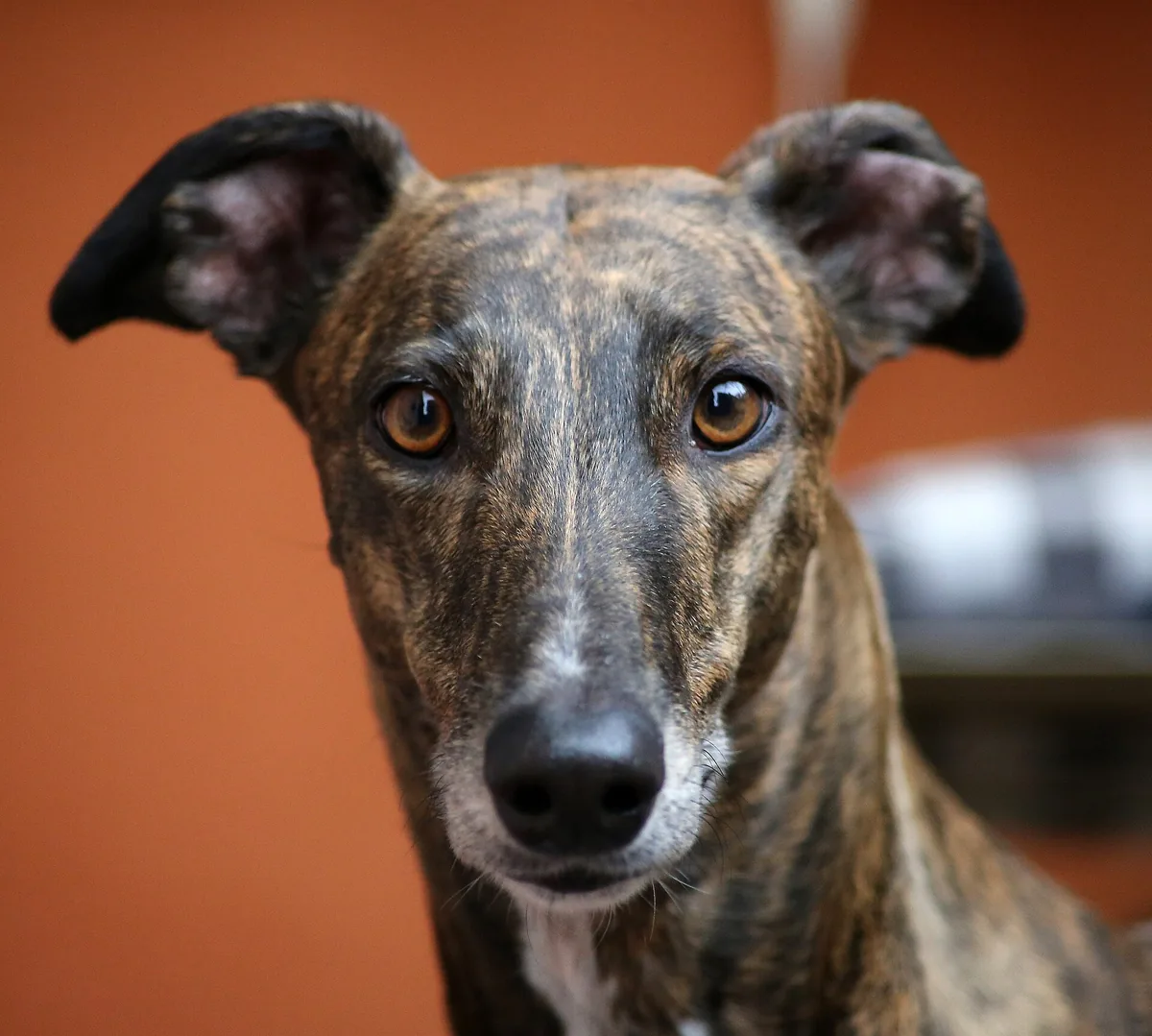
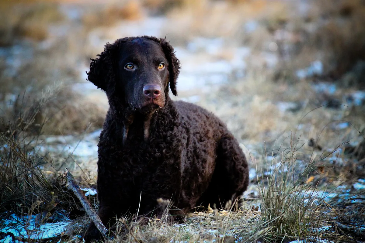
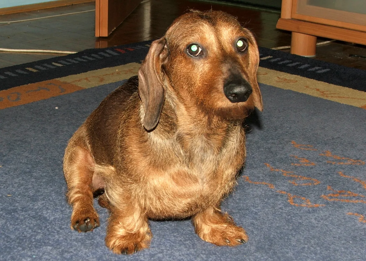
Introductory cards, one per system. The organs are drawn in the atlas’s “Organs” layer, with positions taken from radiology and dissection labs; the path of a scent is in “Journey of a scent”.
The mouth, esophagus, stomach, liver, pancreas, intestines, rectum and anus.
The large and small airways and the lungs; gas exchange takes place in the alveoli.
The heart, with four chambers, and the blood vessels: the arteries and the veins.
The brain, the spinal cord and the nerves throughout the body; it coordinates the organism and the senses. The scent pathway: olfactory nerve → olfactory bulb → piriform lobe → amygdala, hippocampus and frontal cortex.
The kidneys, ureters, urinary bladder and urethra.
Male: the testes and the genital tract. Female: the ovaries, oviducts and uterus. Two distinct anatomies.
Tissues that release hormones into the blood, for example the thyroid and the pituitary.
The thymus and the bone marrow produce the white blood cells; the spleen and the lymph nodes trap microorganisms.
Heart rate
70-120 beats/min
small dogs faster than large ones; in triage 60-120 for large dogs
Respiratory rate
18-34 breaths/min
at rest
Body temperature
38.3-39.2 °C
average
Tables from the MSD Veterinary Manual (after Dukes' Physiology). The reference values do not replace a consultation; a value outside the range is discussed with the veterinarian.
Resources
Worksheets and cards to print, presentation mode for the classroom, the question bank with answers, the full list of structures and the glossary.
For teachers and instructors
Presentation mode puts the drawing across the full width of the screen, with a set of questions for the class (the answer is shown on request). The worksheets print with the answer key on a separate sheet. Nothing requires an account or student names.
105 verified questions, with answer and explanation. Open a question to see the answer; in the lessons, the same questions come without the answer in sight.
Answer: The highest point of the trunk, between the shoulder blades
The withers are the interscapular region, the highest point of the trunk; the dog's height is measured from here.
Answer: Withers · show in the atlas
The withers sit just behind the neck, above the shoulder blades, not in the middle of the back.
Answer: Hock · show in the atlas
The hock is the lower joint of the hind limb, the one that bends backward; the stifle is higher up, near the flank.
Answer: Four: two forelimbs and two hind limbs
The dog is a quadruped: two thoracic (fore) limbs and two pelvic (hind) limbs.
Answer: 7
The dog's vertebral formula is 7 cervical, 13 thoracic, 7 lumbar, 3 sacral and 20-23 caudal.
Answer: 13
13 pairs: 9 sternal and 4 asternal; the last pair is usually floating.
Answer: It is a rudiment of about 1 cm, not connected to the skeleton
In the dog the clavicle is rudimentary and does not articulate with the skeleton; the forelimb is attached to the trunk only by muscles and ligaments.
Answer: Scapula (shoulder blade) · show in the atlas
The scapula is the flat, triangular bone on the side of the thorax, below the withers.
Answer: the femur, tibia and patella
The stifle comprises the femorotibial and femoropatellar joints; the tibia and tarsus form the hock.
Answer: the ankle
The hock is the tarsal joint, the anatomical homolog of the ankle; its point is the calcaneus, that is, the “heel”.
Answer: Stifle (knee) · show in the atlas
The stifle is high up, at the boundary between the thigh and the lower leg, close to the line of the abdomen.
Answer: the wrist
The carpus is the joint between the forearm and the paw, the equivalent of the wrist; it has seven bones.
Answer: 42
42 permanent teeth: 12 incisors, 4 canines, 16 premolars and 10 molars.
Answer: 28
The deciduous dentition has 28 teeth: on each half of the upper and lower jaw, 3 incisors, 1 canine and 3 premolars.
Answer: Molars
The molars and the first premolar have no corresponding deciduous teeth; they erupt directly as permanent teeth.
Answer: may still need treatment if it causes traumatic contact with the gums or the palate
The conformation a standard calls for and dental health are different things: a type of bite considered normal in some breeds does not need treatment unless it produces a traumatic occlusion.
Answer: four beats, each limb striking the ground in turn
At the walk the limbs strike the ground in succession, and alternately two and three limbs are on the ground; diagonal pairs moving together define the trot, not the walk.
Answer: the right forelimb with the left hind limb, then the reverse
The trot is a two-beat gait with diagonal pairs; the hind limb of the diagonal touches the ground slightly before the forelimb.
Answer: the pairs of limbs on the same side
At the pace, the two lateral limbs (on the same side) bear the weight alternately; in dogs it often appears with fatigue or in untrained animals.
Answer: the gallop
The gallop is a four-beat gait with phases of suspension; the rotary variant, with two suspensions, is the fastest.
Answer: from the ground to the highest point of the shoulder blades (withers)
The measurement is taken at the highest point of the shoulder blades, with the dog standing square on a flat, firm surface.
Answer: from the point of the shoulder to the point of the buttock
The FCI model standard defines body length from the point of the shoulder to the point of the buttock; back length (withers to base of tail) is a different measurement.
Answer: does not necessarily coincide with the end of growth; the growth plates close later
Growth continues after the onset of sexual maturity; the skeleton matures at 8-12 months in small and medium breeds and at 10-16 months in large and giant breeds. The two moments do not coincide.
Answer: has one primary hair and several secondary hairs emerging through the same pore
The dog's follicles are compound: a central hair surrounded by 3-15 secondary hairs, all emerging through the same pore. Puppies are born with simple follicles, which become compound.
Answer: the calcaneus
The calcaneus, with its tuberosity, forms the point of the hock; the common calcaneal tendon attaches to it.
Answer: the olecranon of the ulna
The olecranon, the upper end of the ulna, forms the prominence of the elbow.
Answer: right hind, left hind, suspension, left fore, right fore, suspension
The rotary gallop has four beats and two suspensions: one after the second hind limb lifts off, the other after the second forelimb lifts off. It is the dog's fastest gait.
Answer: three beats: one hind limb, a diagonal pair, then the leading forelimb
The canter sequence is 1-2-1; when the diagonal breaks up (the hind limb touches the ground before the forelimb), the canter becomes a transverse gallop.
Answer: about 12-13 %
Craven et al. (2010) measured about 12-13 % with the nostril relaxed; the rest follows the respiratory path toward the pharynx. Nostril movement can raise the percentage.
Answer: down and to the sides, as a jet, because of the shape of the nasal vestibule
The ventrolateral jet stirs up odors from the ground without scattering the sample the dog is about to inhale.
Answer: the nasopalatine duct, which opens behind the upper incisors
The vomeronasal organ sits above the palate, along the nasal septum, and detects low-volatility chemical signals, including pheromones.
Answer: right
Positively charged stimuli (the owner) produce a wider swing to the right; an unfamiliar dog with agonistic behavior, to the left (Quaranta et al. 2007, as cited in the authors' review).
Answer: intense acute stress, including rises in cortisol
Unpredictable stimuli (sudden noises, shocks, a falling bag) produced cortisol responses and a very low posture.
Answer: it occurs most often after a pause and restarts play
In adult dogs and puppies, the bow restarted play after a pause; it did not serve to clarify ambiguous bites. In wolf puppies it did not restart play.
Answer: heightened arousal: it can be fear, surprise, aggression or stress
The reflex appears in several contexts of heightened arousal; it is not a specific sign of aggression.
Answer: is dichromatic: two types of cones, with peaks at about 429 and 555 nm
Neitz, Geist and Jacobs (1989) showed through behavioral tests that the dog has dichromatic vision.
Answer: normally bears no weight and exists only on the forelimbs
It sits on the back of the forelimb, above the accessory carpal bone; the dog walks on its toes.
Answer: 4-7 sniffs per second, regardless of size
Sniffing frequency does not depend on body size; airflow rate and volume do.
Answer: the rudimentary first digit, on the inside of the paw, above the weight-bearing toes
The dewclaw is digit I (the pollex), medial and non-weight-bearing; on the hind limbs it appears only occasionally, and some standards call for double hind dewclaws.
Answer: as a horny sheath over the ungual process of the last phalanx
The ungual process is the cone-shaped end of the last phalanx; the root of the claw sits under the ungual crest, from which it grows.
Answer: the serratus ventralis muscle, attached to the ribs and to the medial face of the scapula
The serratus ventralis carries the weight of the trunk, because the forelimb has no bony articulation with the thorax.
Answer: extends the elbow; its long head also flexes the shoulder
It attaches to the olecranon, the point of the elbow, and is the most powerful muscle of the forelimb; in the dog it actually has four heads.
Answer: the gastrocnemius and the superficial digital flexor, with contributions from the biceps femoris, semitendinosus and gracilis
Because of these contributions, a dog could stand even with the gastrocnemius and the superficial digital flexor paralyzed.
Answer: the diaphragm
The diaphragm, a muscular dome that separates the thorax from the abdomen, is the main muscle of inspiration; the muscles of the thoracic wall help, especially during exertion. The external oblique compresses the abdomen and assists expiration.
Answer: extend the hip: they are the engine of propulsion, the quadruped's “rear-wheel drive”
Running quadrupeds are described as “rear-wheel drive”: the torque generated at the hip pushes the center of gravity forward; in the greyhound these extensors are the largest and most powerful muscles.
Answer: toward the nose; on the head it replaces the term “cranial”
On the head, “cranial” makes no sense (the cranium is at the back of the head), so “rostral” is used (Latin rostrum = beak); “caudal” remains toward the tail, “dorsal” toward the back, “ventral” toward the belly.
Answer: skull width × 100 / skull length
Brachycephalic breeds have a high index (short, wide skull), dolichocephalic breeds a low one; numerical thresholds per breed do not appear in any peer-reviewed source.
Answer: 70-120 beats per minute, faster in small dogs
Values from the MSD tables: 70-120 beats/min (small dogs faster than large ones), respiration 18-34/min, temperature 38.3-39.2 °C. They are guide values, not a diagnostic tool.
Answer: the quadriceps femoris
The quadriceps femoris, with four heads attached to the patella and, through the patellar ligament, to the tibia, extends the stifle; the triceps extends the elbow, and the diaphragm is the main muscle of inspiration.
Answer: between thoracic vertebrae 3 and 8, with its base at the level of the carina
The heart usually sits between T3 and T8, with the middle of its base at the level of the carina (intercostal space 5-6) and its apex on the sternum.
Answer: Right kidney · show in the atlas
The right kidney usually sits more cranially (roughly T13-L2) than the left one (L1-L3); that is why it is drawn behind, with a dashed outline.
Answer: on the left, along the greater curvature of the stomach
The spleen sits on the left abdominal wall, cranially and ventrally, parallel to the greater curvature of the stomach, in the greater omentum.
Answer: the aorta and the esophagus, each through its own hiatus
The crura of the diaphragm attach to the lumbar vertebrae; the aorta passes through the aortic hiatus, the esophagus through the esophageal hiatus.
Answer: immediately caudal to the diaphragm, entirely within the costal arch
The liver lies against the caudal face of the diaphragm and, normally, is seen entirely within the costal arch.
Answer: Stomach · show in the atlas
The stomach sits caudal to the liver, on the left, with its long axis perpendicular to the spine; the spleen accompanies it along the greater curvature.
Answer: it is large in puppies and gradually shrinks in the adult
The volume of the thymus depends on age: large in young animals, it gradually decreases with aging.
Answer: by about 10-17 %
FCI standard no. 166: “the length of the trunk exceeds the height at the withers by about 10-17 %”; the height of males is 60-65 cm.
Answer: the Belgian Shepherd
FCI standard no. 15 says the Belgian Shepherd can be inscribed in a square; the Dachshund's body is 1.7-1.8 times as long as it is tall, the Bloodhound's 10 : 9.
Answer: Head · show in the atlas
The head is the front part of the body, from the tip of the nose to the nape, ears included.
Answer: Tail · show in the atlas
The tail is the continuation of the spine, from the croup downward.
Answer: Ear · show in the atlas
The ear you see is the pinna, on the top of the head, behind the eyes.
Answer: Nose (nose leather) · show in the atlas
The nose leather is the tip of the nose: the moist, hairless skin with the two nostrils through which air enters.
Answer: Muzzle · show in the atlas
The muzzle is the part of the head in front of the eyes: the nose, the jaws and the lips; it ends at the nose leather.
Answer: Neck · show in the atlas
The neck joins the head to the trunk, between the head and the withers.
Answer: Back · show in the atlas
The back is the upper part of the trunk, from the withers to the loin.
Answer: Croup · show in the atlas
The croup is the region above the pelvis, from the front edge of the pelvis to the base of the tail.
Answer: Elbow · show in the atlas
The elbow is the joint between the upper arm and the forearm, on the front leg, below the shoulder.
Answer: Forepaw · show in the atlas
The front paw comprises the metacarpus, the toes and the pads; the dog walks on its toes.
Answer: Hind paw · show in the atlas
The hind paw comprises the metatarsus, the toes and the pads; here too the dog stands on its toes.
Answer: Carpus (wrist) · show in the atlas
The carpus is the joint of the front leg between the forearm and the paw; it corresponds to the human wrist.
Answer: Skull · show in the atlas
The skull is formed by the bones of the head, without the lower jaw; it protects the brain and the sense organs.
Answer: Ribs · show in the atlas
The ribs are curved bones that form the sides of the chest; the dog usually has thirteen pairs.
Answer: Femur · show in the atlas
The femur is the thigh bone, in the hind leg, between the hip and the stifle.
Answer: Pelvis (hip bone) · show in the atlas
The pelvis is formed by the two hip bones, below the croup, between the sacrum and the femur; the head of the femur fits into its socket.
Answer: Mandible · show in the atlas
The mandible is the bone of the lower jaw: it carries the lower teeth and connects to the skull at the jaw joint.
Answer: Heart · show in the atlas
The heart is the four-chambered organ through which the blood circulates; it sits in the central part of the chest.
Answer: Lungs · show in the atlas
The lungs fill the chest cavity, from the thoracic inlet to the diaphragm; the drawing shows the left lung.
Answer: Liver · show in the atlas
The liver lies against the rear face of the diaphragm, inside the arch of the ribs.
Answer: Masseter · show in the atlas
The masseter is the cheek muscle, between the cheekbone and the lower jaw; it raises the mandible, that is, it closes the mouth.
Answer: Diaphragm · show in the atlas
The diaphragm is a large, flat, dome-shaped muscle that separates the chest from the abdomen; it is the main muscle of breathing.
Answer: Triceps brachii · show in the atlas
The triceps sits on the back of the upper arm, from the shoulder blade and the arm bone to the point of the elbow; it extends the elbow.
Answer: the faint warmth of an object warmer than the air, from more than a meter away
Three trained dogs chose, from 1.6 m, a surface 11–13 °C warmer than the air; in 13 dogs scanned with fMRI, the warm stimulus activated the somatosensory cortex. The mechanism is not understood (Bálint 2020).
Answer: the right nostril
Siniscalchi et al. (2011): with new, non-threatening smells, dogs use the right nostril first and switch to the left on repeated presentations; with adrenaline they stay on the right.
Answer: because it cannot pant and sniff at the same time
A dog either sniffs or pants; after 20 minutes of running, 6 detection dogs detected less and searched longer (Gazit and Terkel 2003).
Answer: from 5 footsteps, but not from 3
Six dogs determined the direction from 5 footsteps, not from 3; without human scent, with only the disturbed ground, they failed.
Answer: 3–5 seconds, over 2–5 footprints
Four German Shepherds decided the direction in 3–5 s, sniffing 2–5 footprints at half speed, and got it right in all 20 tests.
Answer: it does not leave: exhalation bypasses the recess and the odor stays on the receptors
Craven (2010): during exhalation no air enters or leaves the olfactory recess; that is why the odor stays on the receptors between two sniffs.
Answer: through the dorsal meatus, a channel under the roof of the nose
The dorsal meatus is the only route that carries odorant-laden air to the olfactory part, bypassing the respiratory turbinates (Craven 2010).
Answer: 1,094, of which 20.3 % pseudogenes
Quignon et al. identified 1,094 genes (minimum estimate), 20.3 % of them pseudogenes; “220 million” refers to cells and comes from a textbook.
Answer: about 0.3 %
Measured on five fixed brains: 0.18 cm³ out of a 58.98 cm³ brain, that is, 0.31 %.
Answer: in the piriform lobe, the olfactory cortex
The lateral olfactory stria carries the signal from the bulb to the piriform lobe, linked to conscious olfaction (UMN); on fMRI, the bulb and the piriform lobes activate in response to smell (Jia 2014).
Answer: the caudate nucleus
The bulb/peduncle reacted the same to all five scents; the caudate responded most to the familiar human, who was not the handler.
Answer: it activated in response to smell mainly in awake dogs, not in anesthetized ones
The bulb and the piriform lobes activated in both states; the frontal cortex and the cerebellum mainly in conscious dogs, which can sniff.
Answer: five tracts from the bulb, including one running directly to the occipital lobe, undocumented in other species
Tracts to the piriform lobe, the limbic system, the entorhinal cortex, the brainstem and the occipital lobe; their functions remain hypotheses, the authors say.
Answer: a bony plate, the lamina transversa
The olfactory recess is excluded from the respiratory path by a bony plate, the lamina transversa (Craven 2010).
Answer: Forehead · show in the atlas
The forehead is the region above the eyes, over the frontal bone.
Answer: Cheek · show in the atlas
The cheek is the lateral wall of the mouth, from the corner of the lips to below the ear.
Answer: Lips · show in the atlas
The lips border the opening of the mouth; in the dog the upper lip covers the lower one.
Answer: Nostrils · show in the atlas
The nostrils are the openings of the nose, in the skin of the nose leather; in the dog they are comma-shaped.
Answer: Nasal bone · show in the atlas
The nasal bones form the roof of the nasal cavity, on the bridge of the nose.
Answer: Maxilla · show in the atlas
The maxilla is the largest bone of the face and carries the upper premolars, molars and canine.
Answer: Cerebellum · show in the atlas
The cerebellum sits behind the hemispheres, above the brainstem.
Answer: Parotid gland · show in the atlas
The parotid sits between the ramus of the mandible, the base of the ear and the masseter.
Answer: Vibrissae (tactile hairs) · show in the atlas
The vibrissae are the thick hairs, with nerves at their base, on the lips, cheeks, chin and above the eyes.
Answer: Tongue · show in the atlas
The tongue sits on the floor of the mouth, between the rami of the mandible.
Below: all structures, in text form · the glossary · frequently asked questions.
Structure cards
The same cards as in the explorer, in text form: name, synonyms, the Latin or English term, location, role and neighboring structures.
The head is the front part of the body, from the tip of the nose to the nape, ears included. It comprises the skull, eyes, ears, muzzle and mouth. Its shape differs a lot from one breed to another, from long muzzles to short ones.
Worth remembering: You can see the bones of the head in the bone layer: the skull and the mandible (lower jaw).
The head comprises the skull, ears, eyes, muzzle and mouth. Its shape and size vary a lot between breeds, from the long muzzles of sighthounds to the short ones of brachycephalic breeds. Inside sit the brain and the sense organs; the bones of the head can be seen in the skeleton layer, as "skull" and "mandible".
When describing the exterior, landmarks such as the forehead, the stop (the transition between forehead and muzzle), the cheeks and the occiput (the upper back part of the head) are used.
The muzzle is the part of the head in front of the eyes: the nose, the jaws and the lips. It starts at the stop, the step between forehead and nose, and ends at the nose leather.
Worth remembering: Inside the muzzle is the nasal cavity, lined with the mucosa through which the dog smells.
The muzzle is the portion of the head in front of the eyes: the nose, the upper jaw and the mandible with the lips. Its length relative to the skull is one of the criteria breed standards use to describe the head. Inside the muzzle is the nasal cavity with the nasal turbinates, covered by the mucosa through which the dog perceives scents.
Each nasal cavity contains three turbinates (nasoturbinate, maxilloturbinate, ethmoturbinate); the olfactory receptors are found mainly on the ethmoturbinates, in the back part of the cavity.
The nose leather is the tip of the nose: the moist, hairless skin with the two nostrils. Through the nostrils, air enters the nasal cavity, where the cells that sense scent are.
Worth remembering: The atlas does not use figures of the "smells X times better" kind: such comparisons say nothing useful about anatomy.
The nose leather is the tip of the nose: the moist, hairless skin with the two nostrils. Through the nostrils, air enters the nasal cavity, where the nasal turbinates and the olfactory epithelium are. Scent is detected through the olfactory epithelium and the olfactory nerves; the vomeronasal organ is an additional pathway for chemical detection.
The atlas does not use multipliers of the "smells X times better" kind: such figures vary enormously between sources and say nothing useful about anatomy.
The eyes sit in the orbits, the bony cavities of the skull, above the stop. Their placement, more to the sides or more to the front, differs between breeds and changes the visual field.
Worth remembering: The retina, the layer at the back of the eye, sends signals to the brain through the optic nerve.
The eyes sit in the orbits, the cavities of the skull that you can see in the skeleton layer as a circle. Their position, more lateral or more frontal, differs between breeds and influences the visual field. The eye is part of the sensory nervous system: the receptors in the retina send signals to the brain through the optic nerve.
The ear you see is the pinna, on the top of the head, behind the eyes. It can be erect, half-raised or drop, depending on the breed. The ear canal and the middle and inner ear are inside the skull.
Worth remembering: Ear position is part of the dog's body language.
The external ear, the pinna, can be erect, semi-erect or drop, depending on the breed. It directs sound toward the ear canal and the middle and inner ear, which lie inside the skull. The pinna is also a communication tool: ear position is part of the dog's body language.
The neck links the head to the trunk, between the head and the withers. Inside are the seven vertebrae of the neck and the muscles that raise, lower and turn the head.
Worth remembering: All breeds have the same number of vertebrae in the neck, seven; only their length differs.
The neck links the head to the trunk. Inside are the seven cervical vertebrae and the muscles that let the dog raise, lower and turn its head. The collar sits on the neck; the length and "arch" of the neck are frequent landmarks in breed standards.
All breeds have the same number of cervical vertebrae, seven; what differs is their length and that of the surrounding muscles.
The withers are the highest part of the trunk, just behind the neck. They lie between the shoulder blades, the bones of the shoulder area. They are the landmark for measuring a dog's height, vertically from the ground.
The withers are the highest point of the trunk, between the shoulder blades. Under the skin lie the spinous processes of the first thoracic vertebrae and the upper edges of the two scapulae. They are the standard landmark for a dog's height: "height at the withers" is measured from here to the ground.
For how to measure correctly, see the module "How to measure a dog": the dog stands square on a flat surface and the measurement is taken at the highest point of the shoulder blades.
The back is the upper part of the trunk, from the withers to the loin. Beneath it lie the vertebrae to which the ribs attach.
Worth remembering: The "topline" is the dog's upper profile seen from the side: withers, back, loin and croup.
The back is the upper part of the trunk, from the withers to the loin. Beneath it lie the thoracic vertebrae, to which the thirteen pairs of ribs attach. In show-ring vocabulary, "back" usually means the region between the withers and the base of the tail, but some standards restrict it to the area above the thorax.
The "topline" is the dog's upper profile seen from the side: withers, back, loin and croup.
The loin is the portion of the back between the last rib and the pelvis. Beneath it are the lumbar vertebrae, which have no ribs, so the area is supported only by muscle.
Worth remembering: Much of the spine's flexibility at the gallop comes from here, when the back arches and stretches.
The loin is the portion of the back between the last rib and the pelvis. Beneath it lie the seven lumbar vertebrae, which have no ribs, so the region is supported only by muscle. Much of the spine's flexibility at the gallop comes from here, when the back arches and stretches.
The croup is the region above the pelvis, toward the tail. It extends from the front edge of the pelvis to the base of the tail.
The croup is the part of the back above the pelvis, from its front edge to the root of the tail. Beneath it lies the sacrum, the three fused vertebrae to which the pelvis attaches. The slope of the croup influences the angle at which the tail is carried and the way the hindlimb pushes.
The thorax is the front part of the trunk, between the front legs and the abdomen. It is the cage formed by vertebrae, ribs and sternum, which houses the heart and lungs.
Worth remembering: Chest depth and circumference are common landmarks in standards and when choosing a harness.
The thorax is the cage formed by the thoracic vertebrae, the thirteen pairs of ribs and the sternum. It houses the heart and lungs. Its depth, measured from the withers to the bottom of the chest, and its circumference are common landmarks in standards and in choosing a harness.
Chest circumference is measured in different places depending on the source (around the deepest part of the chest, just behind the shoulders, or behind the withers), so when you compare figures, check where each one was measured.
The abdomen is the lower and rear part of the trunk, between the last rib and the pelvis. It has no bones on its sides: its wall is made of muscle.
Worth remembering: Inside sit the stomach, intestines, liver, kidneys, spleen and urinary bladder.
The abdomen is the portion of the trunk behind the ribs, as far as the pelvis. It has no bones on its sides: its wall is muscular. Inside sit the stomach, intestines, liver, kidneys, spleen and urinary bladder. The underline, from the chest toward the flank, "rises" more or less depending on the breed.
The tail is the continuation of the vertebral column, from the croup downward. It is made of vertebrae that get smaller toward the tip, and their number differs from one dog to another.
Worth remembering: The tail helps with balance in turns and communicates through position and movement.
The tail is the continuation of the vertebral column: usually between twenty and twenty-three caudal vertebrae, getting smaller toward the tip. The number varies between individuals. The tail helps with balance in turns and is a communication tool, through position and movement.
Tail shape and length differ a lot between breeds, and the number of vertebrae can differ even between dogs of the same breed.
The forechest is the front part of the trunk, below the neck, between the front legs. Beneath it lie the front end of the sternum and the shoulder joints.
Worth remembering: How far the chest "projects" forward is a common landmark in breed standards.
The forechest is the front part of the trunk, between the two forelimbs and below the base of the neck. Beneath it lie the front end of the sternum, the manubrium, and the shoulder joints. How far the chest "projects" forward of the point of the shoulder is a common landmark in standards.
The shoulder is the upper part of the front leg, on the side of the chest. It comprises the shoulder blade, the flat bone of the shoulder, and the muscles that attach it to the trunk.
Worth remembering: The dog has no functional collarbone: the front leg is attached to the body only by muscles and ligaments.
The shoulder is the upper region of the forelimb: the shoulder blade (scapula) and the muscles that attach it to the trunk. The dog has no functional collarbone, so the forelimb is attached to the body only by muscles and ligaments, not by a bony joint. The shoulder joint is between the scapula and the humerus.
The angle between the shoulder blade and the upper arm (the "scapulohumeral angle") is one of the conformation landmarks most often discussed in working dogs.
The upper arm is the segment of the front leg between the shoulder and the elbow, on the side of the chest. Its bone is the humerus, covered by the muscles that flex and extend the elbow.
Worth remembering: In the standing dog, the upper arm slopes backward, and the elbow comes about level with the bottom line of the chest.
The upper arm is the segment between the shoulder joint and the elbow. Its bone is the humerus, covered by the muscles that extend and flex the elbow. In the standing dog, the upper arm slopes backward, and the elbow comes roughly level with the bottom line of the chest.
The elbow is the joint between the upper arm and the forearm. You find it on the front leg, below the shoulder.
Worth remembering: The point of the elbow is an extension of one of the two bones of the forearm.
The elbow is the joint between the upper arm and the forearm, formed by the humerus, radius and ulna. The point of the elbow, the bump you can easily feel with your fingers, is the olecranon, the upper end of the ulna. The elbow bends in a single plane, forward and backward, like a hinge.
The forearm is the segment between the elbow and the wrist of the front leg. It has two bones lying side by side: the radius in front and the ulna behind.
Worth remembering: In the standing dog, the forearm is nearly vertical.
The forearm is the segment between the elbow and the wrist. It has two bones, the radius and the ulna, lying side by side: the ulna sits behind at the elbow and on the outer side at the wrist, the radius in front. In the standing dog, the forearm is nearly vertical.
The carpus is the wrist of the front leg, between the forearm and the paw. It corresponds to the human wrist and is made of seven small bones, in two rows.
Worth remembering: On the back of the carpus is the carpal pad, the only pad that does not touch the ground when walking.
The carpus is the wrist of the forelimb, between the forearm and the paw: the counterpart of the human wrist. It is made of seven small bones, arranged in two rows. On the back of the carpus is the carpal pad, the only pad that does not touch the ground when walking.
The front paw comprises the metacarpus (the part between the wrist and the toes), the toes and the pads. The dog walks on its toes, with the metacarpus raised off the ground.
Worth remembering: Four toes bear the weight; the fifth, the dewclaw on the inner side, is slender and does not touch the ground.
The forepaw comprises the metacarpus, the toes and the pads. The dog is digitigrade: it walks on its toes, with the metacarpus raised off the ground. Four toes bear the weight, each with a pad and a claw; the fifth, the dewclaw (digit I, on the inner side), is slender and takes no part in support.
The pads are made of connective and fatty tissue, which cushions, and of a thick, horny epidermis. On the forepaw a dog has one pad on each of digits II-V, a common metacarpal pad and the carpal pad.
The thigh is the upper part of the hind leg, from the hip to the stifle. Its bone is the femur, surrounded by the dog's most powerful muscles.
Worth remembering: The hind legs are the "engine" that pushes the body forward.
The thigh is the upper segment of the hindlimb, from the hip joint to the stifle. Its bone is the femur, surrounded by the most powerful muscles of the dog: the hindlimbs are the "engine" of locomotion, the ones that push the body forward.
Unlike the forelimb, the hindlimb attaches to the spine through a bony joint, the sacroiliac joint, via the pelvis.
The stifle is the joint at the lower end of the thigh, on the hind leg. It sits above the hock, the joint that corresponds to the human ankle.
Worth remembering: The patella (kneecap) is one of the bones that form the stifle.
The dog's knee, called the stifle, is the joint between the thigh and the lower thigh: femur, tibia and patella. It sits higher than you would think, near the flank, and bends forward, like the human knee. Don't confuse it with the hock, the joint below it, which bends backward.
Some sources wrongly call it the "femorotibiotarsal" joint; correctly, the stifle is formed by the femorotibial and femoropatellar joints, without the tarsal bones.
The lower thigh is the segment of the hind leg between the stifle and the hock. It has two bones: the tibia, thicker, in front, and the fibula, slender, on the outer side.
Worth remembering: In the standing dog the lower thigh slopes backward; its length relative to the thigh gives the "angulation" of the hind leg.
The lower thigh is the segment of the hindlimb between the stifle and the hock. It has two bones: the tibia, thicker, in front, and the fibula, slender, on the outer side. In the standing dog the lower thigh slopes backward, and its length relative to the thigh gives the "angulation" of the hindlimb.
The hock is the joint of the hind leg that corresponds to the human ankle. It sits below the stifle, above the paw.
Worth remembering: The point of the hock is a projection of the calcaneus, one of the bones of this joint.
The hock is the joint between the lower thigh and the hind paw, the counterpart of the human ankle. It bends backward, the opposite of the stifle, which is why it looks like a "reversed knee". The point of the hock, the bump at the back, is the calcaneus, the bone to which the Achilles tendon attaches.
The tarsus has seven bones. "Hock" comes from show-ring and horse vocabulary; in anatomy the region is called the tarsus.
The hind paw comprises the metatarsus (the part between the hock and the toes), the toes and the pads. As in front, the dog stands on its toes, with the metatarsus raised off the ground.
Worth remembering: The hind dewclaw is absent in many dogs.
The hind paw comprises the metatarsus, the toes and the pads: a common metatarsal pad and one pad on each of the four toes that touch the ground. As in front, the dog stands on its toes, with the metatarsus raised. The hind dewclaw is absent in many dogs.
The forehead is the part of the head above the eyes, on the frontal bone. In the dog it has a small hollow in the middle and slopes down toward the nose.
Worth remembering: The width and shape of the forehead depend on the breed; under it is the frontal bone with the frontal sinus.
The forehead is the region over the frontal bone, between the eyes and the crown, continued toward the nose by the bridge of the nose. In the dog, the frontal parts of the two bones form a central depression and slope forward, toward the stop. Under the skin lie the frontalis muscle, weak in the dog, and the bundles of the levator nasolabialis, which start here toward the upper lip.
The stop is the step between the forehead and the nose, between the eyes. The word comes from the world of dog shows, not from anatomy.
Worth remembering: Pronounced in short-muzzled breeds, almost absent in sighthounds; the FCI standards describe it for each breed.
The stop is the depression between the eyes, where the nasal bones meet the skull. It is a term from the show-ring vocabulary, defined by the Kennel Club glossary, not from anatomical nomenclature: the anatomy pages consulted do not use it. Anatomically, the area corresponds to the junction of the nasal bones with the frontal bone, where the frontals form a central depression.
The cheek is the lateral wall of the mouth. The back part is firm, over the masseter muscle; the front part is soft and moves together with the lips.
Worth remembering: Under the cheek run the parotid duct and the branches of the facial nerve, which moves the face.
The cheek is the lateral wall of the mouth. It has a masseteric part, firm, over the masseter muscle, and a buccal part, mobile, toward the corner of the mouth, whose foundation is the buccinator muscle. In the dog the cheeks are loose and roomy, with a smooth, often pigmented mucosa. The duct of the parotid gland crosses the cheek and opens opposite the upper teeth.
The lips are the soft edges of the mouth. In the dog they are thin, move a lot and carry tactile hairs; the upper lip is larger and covers the lower one.
Worth remembering: Don't take “pendulous” lips for a sign of disease: in some breeds that is simply how they are.
The lips border the opening of the mouth. In the dog they are thin and mobile, with numerous tactile hairs; the upper lip is larger than the lower one, which it covers, and the edges of the lower lip are flaccid and serrated. “Flews” is the show-ring word for the pendulous upper lips of some breeds. In their thickness lies the orbicularis oris, supplied by the facial nerve.
The chin is the part below the lower lip, at the tip of the jaw. In the dog it sticks out far less than in humans.
Worth remembering: The tactile hairs on the chin are part of the vibrissa system described in the dog.
The chin is the fleshy prominence below the lower lip, at the tip of the mandible. In animals it is far less marked than in humans. From here, on the inner side, start the muscles that pull the tongue forward: the genioglossus, “from chin to tongue”, and the geniohyoideus. The mental tactile hairs also sit on the chin.
The nostrils are the openings of the nose. In the dog they are comma-shaped, with the wide part toward the middle and the narrow tail toward the side and backward.
Worth remembering: Each nostril takes a separate sample of air; see “Journey of a Scent”.
The nostrils are the external openings of the nasal cavity, set in the planum nasale, the bare skin of the nose leather. In the dog each nostril is comma-shaped, with the wide part next to the septum and the narrow tail pointing backward and outward, into a groove, the alar groove, barely marked in carnivores. The upper edge, the wing of the nostril, is convex and mobile; the levator nasolabialis pulls on it.
Vibrissae are the “whiskers” and the other thick hairs on the face: above the eyes, on the lips, on the cheeks, on the chin. They are organs of touch, not simple hairs.
Worth remembering: Don't trim them: they are sensory hairs, with nerves at the base, and the dog uses them to explore.
Vibrissae are thick, richly innervated hairs that serve the sense of touch. In the dog six groups are confirmed: above the eyes, on the upper lip (the whiskers), on the lower lip, on the chin, on the cheeks and in the zygomatic region, plus microvibrissae on the lips. Touching the ones above the eyes or the whiskers triggers a reflex blink; the dog pushes its whiskers forward when exploring an object.
The eyelids are the skin that closes the eye. The dog also has a third eyelid, in the corner toward the nose, which slides across the eye when the eye is drawn back.
Worth remembering: A third eyelid that stays visible for a long time is a reason to visit the vet, not to diagnose at home.
The eyelids are mobile folds that protect the eye; their middle layer contains the fibers of the orbicularis oculi. In carnivores, the lower eyelid has no cilia. In the corner toward the nose sits the third eyelid, the nictitating membrane, supported by a T-shaped cartilage: its movement is passive, it is pushed across the cornea when the retractor muscle pulls the eyeball back; humans have neither a third eyelid nor this muscle.
The temporalis is the most massive muscle of the head. It sits on the sides of the skull, from above the eye to the nape, and attaches to the lower jaw.
Worth remembering: It closes the mouth, so it gives the bite its force, and it guards the jaw joint.
The most massive muscle of the head in carnivores, thick and elastic. It arises from the temporal fossa (and from the sagittal crest) and inserts by a tendinous sheet on the coronoid process of the mandible. It closes the mouth, so it bites, and it guards the temporomandibular joint against forward luxation.
The masseter is the cheek muscle, between the zygomatic arch (the cheekbone) and the lower jaw. It raises the mandible, that is, closes the mouth.
Worth remembering: Together with the temporalis it gives the bite its force; the mouth is opened by another muscle, the digastricus, on the underside of the jaw.
It arises from the lower edge of the zygomatic arch and inserts on the lateral face of the mandible (the masseteric fossa). It raises the mandible, that is, closes the mouth; together with the temporalis it gives the bite its force. The only masticatory muscle that opens the mouth is the digastricus, on the underside of the jaw.
The brachiocephalicus is the band of muscle on the front of the neck, from the head to the bone of the upper arm. It has two parts, separated where the remnant of the collarbone lies.
Worth remembering: It pulls the front leg forward; when the leg is planted on the ground, it extends the neck or turns the head.
It links the arm to the head and neck and has two parts, separated by the clavicular intersection, where the vestige of the clavicle lies: the cleidobrachialis, attached to the humerus, and the cleidocephalicus, attached to the nape and the mastoid process. It pulls the limb forward; when the limb is fixed under the body's weight, it extends the neck or turns the head.
The sternocephalicus is the muscle on the underside of the neck, from the sternum to the head. It bends the head and neck downward.
Worth remembering: When it works on one side only, it tilts and rotates the head toward that side.
It arises from the manubrium of the sternum and runs up the underside of the neck to the mastoid process and the occipital region of the skull. It flexes the head and neck; when only one side contracts, it tilts and rotates the head toward that side.
The trapezius is a broad, triangular muscle on the withers and the upper part of the chest, above the shoulder blade. It arises from the midline of the back and attaches to the shoulder blade.
Worth remembering: It raises the front leg and pulls it forward.
A broad, triangular muscle with a cervical part and a thoracic part, separated by an aponeurotic zone. It arises from the dorsal midline, from the third cervical vertebra to the ninth thoracic, and inserts on the spine of the scapula. It raises the forelimb and pulls it forward.
The latissimus dorsi is a broad sheet of muscle on the sides of the chest, behind the shoulder blade, from the loin to the upper arm.
Worth remembering: It pulls the front leg backward; in the greyhound it is among the muscles that do most of the work of acceleration.
A broad muscular sheet that arises from the thoracolumbar fascia (the lumbar and last thoracic vertebrae, the last ribs) and converges on the teres major tuberosity of the humerus. It pulls the free limb backward and brakes its advance; in the greyhound it is among the muscles that produce most of the mechanical work of acceleration.
The deltoideus sits over the shoulder joint, from the shoulder blade to the bone of the upper arm. In the dog it has two parts.
Worth remembering: It bends the shoulder and raises the bone of the upper arm.
In the dog it has two parts, scapular and acromial. It arises from the spine and the acromion of the scapula and inserts on the deltoid tuberosity of the humerus. It flexes the shoulder joint and raises the humerus.
The triceps is the strongest muscle of the front leg. It sits on the back of the upper arm, from the shoulder blade and the arm bone to the point of the elbow.
Worth remembering: It extends the elbow at every stride; in the dog it has four heads, although the name says “three”.
The strongest muscle of the forelimb. The long head arises from the caudal border of the scapula, the other heads from the humerus (in the dog there are four, although the name says “three”); all insert on the olecranon, the point of the elbow. It extends the elbow at every stride; the long head also flexes the shoulder.
The biceps brachii sits on the front of the upper arm, mostly on the inner face; from the side only its edge shows. In the dog it has a single head.
Worth remembering: It bends the elbow and extends the shoulder.
In the dog it has a single head. It arises from the supraglenoid tubercle of the scapula and inserts by a branched tendon on the tuberosities of the radius and ulna. It flexes the elbow and extends the shoulder.
The pectorals are the chest muscles, from the sternum to the bone of the upper arm; from the side their edge shows below the chest. They fasten the front leg to the trunk.
Worth remembering: The deep pectoral pulls the trunk forward over the leg planted on the ground; in the greyhound it is the heaviest muscle of the front leg.
Extrinsic muscles that fasten the limb to the sternum. The superficial pectorals insert on the crest of the greater tubercle of the humerus and support the limb, pulling it inward; the deep pectoral, the heaviest muscle of the forelimb in the greyhound, pulls the trunk forward over the supporting limb and extends the shoulder.
The serratus ventralis sits on the sides of the chest, below the shoulder blade, as a band with “teeth” attached to the ribs. It attaches to the inner face of the shoulder blade.
Worth remembering: It is the “sling” that carries the weight of the trunk between the two shoulder blades, because the front leg has no bony link to the chest.
It arises by digitations from the ribs and the cervical vertebrae and inserts on the medial face of the scapula. Since the forelimb has no bony joint with the thorax, this muscle is the “sling” that carries the weight of the trunk between the two shoulder blades; it also helps with inspiration.
The external oblique is the muscle of the lateral wall of the abdomen, from the last ribs to the midline of the belly. Its fibers run obliquely downward and backward.
Worth remembering: It presses on the organs in the abdomen, which helps with expiration, and supports the wall of the belly.
Its fibers arise from the caudal ribs and the thoracolumbar fascia and run obliquely downward and backward, to an aponeurosis attached to the linea alba. It compresses the abdominal viscera (the “abdominal press”), which helps with expiration, and supports the abdominal wall.
The epaxial muscles lie along the spine, above it, from the pelvis to the head; in the drawing they are the band below the trapezius and the latissimus dorsi. The longest of them, the longissimus, runs from the pelvis to the head.
Worth remembering: They extend the spine; at the gallop, the bending and straightening of the back greatly lengthen the stride.
The epaxial muscles lie dorsal to the spine and extend it; the largest of them, the longissimus, runs from the pelvis to the head and is the longest muscle in the body. At the gallop, flexion and extension of the spine greatly lengthen the stride: these muscles are part of the engine of running.
The gluteus medius sits on the croup, above the hip, on the wing of the pelvis. It attaches to the femur, at the bump below the hip.
Worth remembering: It extends the hip, so it pushes the body forward.
It arises from the gluteal surface and the crest of the ilium and inserts on the greater trochanter of the femur. It extends the hip joint, so it pushes the body forward; it also tends to rotate the femur with the stifle inward, a tendency counterbalanced by the caudal hip muscles.
The gluteus superficialis sits on the croup, behind the gluteus medius, toward the tail. It arises from the sacrum and attaches to the femur.
Worth remembering: It extends the hip joint.
It arises from the gluteal fascia, from the sacrum and from the sacrotuberous ligament, and inserts on the third trochanter of the femur. It extends the hip joint.
The biceps femoris is the largest of the muscles at the back of the thigh. It sits on the lateral and rear part of the thigh, from the pelvis to the stifle and hock.
Worth remembering: It supports the body when the leg is on the ground and is, together with the adductor, the strongest extensor of the hip.
The largest of the muscles at the back of the thigh (the “hamstrings”). It arises from the ischiatic tuberosity and the sacrotuberous ligament and inserts widely on the fascia lata and crural fascia, on the tibia and, through the calcaneal tendon, on the calcaneus. It supports the body in the stance phase, flexes the stifle at the start of the swing and extends the hock; together with the adductor it is the strongest extensor of the hip.
The semitendinosus sits on the rear edge of the thigh, from the pelvis to the lower leg and the point of the hock.
Worth remembering: It extends the hip and hock: it is part of the engine of forward drive.
It arises from the lateral angle of the ischiatic tuberosity, between the biceps femoris and the semimembranosus, and inserts on the medial face of the tibia and on the calcaneus. It extends the hip and hock and flexes the stifle of the free limb; it is one of the main extensors of the hip, so part of the engine of propulsion.
The quadriceps is the muscle at the front of the thigh, from the pelvis and femur to the kneecap. It has four heads; all attach to the kneecap, which is linked to the tibia by a ligament.
Worth remembering: It extends the stifle; one of its heads also bends the hip.
Four heads: the rectus femoris arises from the ilium, the three vasti from the femur. All insert on the patella, and the patella is linked to the tibial tuberosity by the patellar ligament. It extends the stifle; the rectus femoris also flexes the hip and is especially powerful in the greyhound.
The gastrocnemius is the muscle at the back of the lower leg, from the femur to the point of the hock, where it attaches through the calcaneal tendon.
Worth remembering: It extends the hock and slightly bends the stifle.
It has two heads, which arise from the lateral and medial supracondylar tuberosities of the femur, and it inserts on the calcaneus through the common calcaneal tendon. It mainly extends the hock and slightly flexes the stifle; its lever arm at the hock stays large over the whole range of motion.
The calcaneal tendon, or Achilles tendon, lies above the point of the hock, on the back of the lower leg. It is not a single tendon but a bundle of the tendons of several muscles.
Worth remembering: Because of this makeup, a dog could still stand even if some of these muscles stopped working.
It is not a single tendon: its core is formed by the tendons of the gastrocnemius and the superficial digital flexor, with contributions from the biceps femoris, semitendinosus and gracilis. That is why a dog could still stand even if the gastrocnemius and the superficial digital flexor were paralyzed.
The diaphragm is a large, flat, dome-shaped muscle inside the trunk. It completely separates the chest cavity from the abdominal cavity; in the drawing it is the dotted line, bulging forward.
Worth remembering: It is the main muscle of inspiration; the chest muscles help, especially during exertion.
A large, flat, dome-shaped muscle that completely separates the thoracic cavity from the abdominal cavity. It arises from the lumbar vertebrae (the crura of the diaphragm), from the ribs, the costal cartilages and the xiphoid process, and inserts on the central tendon. It is the main muscle of inspiration; the muscles of the thoracic wall help, especially during exertion.
The cupola, the highest part of the dome, is asymmetrical in the dog: on the left it reaches the level of the sixth rib, on the right one intercostal space further back; the esophageal hiatus lies ventral to thoracic vertebra 12, and the costal part attaches to the cartilage of the ninth rib, to cartilages 10-11 slightly below the junction with the rib, to rib 12 at its ventral end and to the last rib below its middle (Sisson, 1914).
The orbicularis oris is the muscle in the thickness of the lips, which closes and purses the mouth. In the dog it is thin and split in two at the middle of the upper lip.
Worth remembering: It is supplied by the facial nerve: when one half of the face “drops”, the lips on that side go slack.
The orbicularis oris lies in the thickness of the lips, around the opening of the mouth, with no attachment to bone. In carnivores, the part in the upper lip is interrupted on the midline by a raphe, and in the dog the muscle is weakly developed. Each half is supplied separately by the facial nerve of its own side. The platysma, the cutaneous muscle of the neck, usually attaches to it.
The levator nasolabialis runs down from the forehead toward the nose and the upper lip. It lifts the lip and pulls on the wing of the nostril: it is one of the muscles the dog moves its nose with.
Worth remembering: There are no “special” nasal muscles in the dog, according to Sisson; the nostril moves through this muscle and through the levator of the upper lip.
The levator nasolabialis starts broadly from the frontal and nasal bones, in the dog also from the upper edge of the maxilla, and runs down under the skin to the upper lip and the wing of the nostril. It lifts the upper lip, pulling it laterally, and pulls on the wing of the nostril. In carnivores it is very broad and without clear limits. Its motor nerve is the dorsal buccal branch of the facial nerve.
The buccinator is the thin muscle in the cheek. It pulls the corner of the mouth backward and pushes food toward the teeth; the name comes from “trumpeter”.
Worth remembering: It is the foundation of the cheek: the soft cheek in front of the masseter is, in fact, the buccinator covered by skin.
The buccinator is the flat muscle of the cheek, stretched parallel to the axis of the mouth; its molar part arises from the maxilla, above the cheek teeth. In the dog it is broad and very thin, with two planes of crossed fibers. It pulls the corner of the mouth backward and laterally and keeps the food between the teeth. It is supplied by the buccal branches of the facial nerve, mainly the dorsal one.
The orbicularis oculi is the ring-shaped muscle in the eyelids, with which the dog blinks and closes its eye.
Worth remembering: The reflex blink when the whiskers or the hairs above the eyes are touched goes through this muscle.
The orbicularis oculi surrounds the opening of the eye; its fibers form the middle, fibromuscular layer of the eyelids and lie next to the tarsus. In the dog, the cutaneous muscle of the face sends a layer toward the lower eyelid that merges with the orbicularis, the malaris muscle. It is supplied by the facial nerve, through the zygomatic branch, the one serving the eyelids and the region of the nose.
The digastricus is the muscle that opens the mouth: it starts behind the ear and pulls the lower jaw down.
Worth remembering: The masseter and the temporalis close the mouth; the digastricus opens it.
The digastricus arises from the paracondylar process of the occipital bone and inserts on the ventromedial edge of the mandible. It opens the mouth: when the masticatory muscles relax, it pulls the mandible down. It has two bellies, with different nerve supply, from the trigeminal and from the facial nerve. In Sisson's old nomenclature it appears as the occipitomandibularis, with the same attachments.
The auricular muscles are the small muscles around the base of the ear, with which the dog turns and raises its ears, each one separately.
Worth remembering: Their exact number does not appear in a verifiable source; the groups do.
The auricular muscles are the small groups of muscles that move the pinna: the rostral group, which contains the scutiform cartilage, a flat plate in front of the ear, and the caudal group. Sisson lists in the dog the scutularis, the anterior auricular muscles, the helicis, the antitragicus and the parotidoauricularis. All are supplied by the facial nerve, which serves the superficial muscles of the head and of the external ear.
The skull is made of the bones of the head, without the lower jaw. It protects the brain and the sense organs and forms the face: the orbits in which the eyes sit, the nasal cavity and the upper jaw with the upper teeth.
Worth remembering: Skull shape differs between long-muzzled breeds, short-muzzled breeds and those in between.
The skull protects the brain and the sense organs and forms the face: the orbits in which the eyes sit, the nasal cavity with the turbinates and the maxilla with the upper teeth. It is made of several bones joined by sutures. The mandible articulates with the temporal bone at the temporomandibular joint.
Skull shape ranges from dolichocephalic breeds (longer longitudinal axis) to brachycephalic ones (short, wide skull, short muzzle, wide zygomatic arches, rounded neurocranium), with mesaticephalic breeds in the middle. The measure used is the cephalic index: skull width × 100 / skull length (from prosthion to inion); numerical thresholds per breed do not exist in a peer-reviewed source.
Palpable landmarks: the zygomatic arch, the external occipital protuberance (the highest point of the skull in the dog), the sagittal crest (variable between breeds), the temporomandibular joint and the tympanic bulla, all selectable in the skeleton layer.
The mandible is the bone of the lower jaw. It carries the lower teeth and connects to the skull at the jaw joint, the one that opens and closes the mouth.
Worth remembering: Its length relative to the upper jaw decides how the lower teeth sit against the upper ones (the occlusion).
The mandible is the bone of the lower jaw. It carries the lower teeth and articulates with the temporal bone of the skull at the temporomandibular joint, the one that allows the mouth to open and close. Its length relative to the maxilla determines how the lower teeth sit against the upper ones, that is, the occlusion.
The cervical vertebrae are the bones of the spine in the neck: seven in all breeds. They carry the head, and the neck muscles attach to them.
Worth remembering: The first two, the atlas and the axis, have special shapes and allow the "yes" and "no" movements of the head.
The cervical spine has seven vertebrae, in all breeds, from the Chihuahua to the Great Dane. The first two, the atlas and the axis, have special shapes and allow the "yes" and "no" movements of the head. The cervical vertebrae carry the head and are the attachment point of the neck muscles.
The dog's vertebral formula: 7 cervical, 13 thoracic, 7 lumbar, 3 sacral and 20-23 caudal; about 4.5% of dogs have an extra lumbar vertebra or a transitional vertebra.
The thoracic vertebrae are the bones of the spine above the chest, from the withers to the last rib. Each of the thirteen carries a pair of ribs.
Worth remembering: Their upward projections, tall at the first vertebrae, form the withers together with the edges of the shoulder blades.
The thirteen thoracic vertebrae each carry a pair of ribs. Their spinous processes, the upward projections, are tallest at the first vertebrae and form the withers together with the edges of the scapulae. Toward the rear, the processes get shorter and the back becomes more flexible.
The lumbar vertebrae are the bones of the spine between the last rib and the sacrum, under the loin. They are the largest vertebrae and have no ribs.
Worth remembering: Their wide lateral projections anchor the powerful muscles of the back.
The seven lumbar vertebrae are the largest vertebrae of the spine and have no ribs. Their wide transverse processes anchor the powerful muscles of the back. The number sometimes varies: in a study of 89 dogs, about 4.5% had an extra lumbar vertebra or a transitional one.
The sacrum is the bone under the croup, between the lumbar vertebrae and the tail. It is made of three vertebrae fused into a single bone.
Worth remembering: The pelvis attaches to it; through this bony link the force of the hind legs reaches the spine and pushes the body forward.
The sacrum is made of the three sacral vertebrae, fused into a single bone. The pelvis attaches to it through the sacroiliac joint: this is the bony link through which the force of the hindlimbs reaches the spine and pushes the body forward. The sacrum continues with the tail vertebrae.
The caudal vertebrae are the bones of the tail. They get smaller toward the tip, where they end up as simple bony nodules; their number differs from one dog to another.
Worth remembering: The first tail vertebrae still have projections and a canal for the nerves; the last ones are just bodies of bone.
The caudal vertebrae form the skeleton of the tail: usually between twenty and twenty-three, getting smaller toward the tip, where they become simple bony nodules. The number differs between dogs. The first tail vertebrae still have processes and a vertebral canal; the last ones are just vertebral bodies.
The ribs are curved bones that form the sides of the chest. The dog normally has thirteen pairs.
Worth remembering: The ribs protect the heart and lungs.
The dog has thirteen pairs of ribs: nine sternal, which reach the sternum through their cartilage, and four asternal, whose cartilages join each other and form the costal arch. The last pair is usually "floating", free at its lower end. The ribs protect the heart and lungs.
The illustration shows the thirteen ribs of the left side; the costal cartilages are not drawn separately.
The sternum is the bone on the bottom line of the chest, between the two rows of ribs. It is made of eight segments joined by cartilage.
Worth remembering: The front end can be felt at the forechest; the rear end sits at the start of the abdomen.
The sternum is the bone on the bottom line of the chest. In the dog it is made of eight sternebrae, bony segments joined by cartilage, which remain separate throughout life. The front end, the manubrium, can be felt at the forechest; the rear end, the xiphoid process, sits at the start of the abdomen.
The scapula is a bone of the shoulder. It lies on the side of the chest, at the upper end of the front leg.
Worth remembering: The muscles around it support the weight of the trunk between the front legs.
The scapula is the flat, triangular bone of the shoulder. It has no joint with the trunk: in the dog the clavicle is a rudiment of about one centimeter, not connected to the skeleton, so the forelimb "hangs" from the body in a sling of muscles and ligaments. With the humerus it forms the shoulder joint.
The serratus ventralis muscle carries the weight of the trunk between the two scapulae; when standing, the forelimbs support about 60% of the body weight.
The humerus is the bone of the upper arm, from the shoulder to the elbow. At the top it connects to the shoulder blade, at the bottom to the two bones of the forearm.
Worth remembering: In the standing dog it slopes backward, which is why the elbow comes under the chest, not under the shoulder.
The humerus is the bone of the upper arm. Its round head articulates with the scapula at the shoulder, and its lower end with the radius and ulna at the elbow. In the standing dog it slopes backward, which is why the elbow comes under the thorax, not under the shoulder. It is one of the strongest bones of the forelimb.
The radius is the front bone of the forearm, from the elbow to the wrist. It carries most of the weight that passes through the forearm.
Worth remembering: It lies alongside the ulna; the two bones rotate slightly relative to each other.
The radius is the front bone of the forearm and the one that carries most of the weight between the elbow and the carpus. It articulates with the humerus above and with the carpal bones below. Together with the ulna it forms a pair of bones lying side by side, with little rotation between them.
The ulna is the rear bone of the forearm, alongside the radius. Its upper end, the olecranon, is the bump we call the point of the elbow.
Worth remembering: The tendon of the triceps, the muscle that extends the elbow at every stride, attaches to the olecranon.
The ulna is the second bone of the forearm. It sits behind the radius at the elbow and on the outer side at the carpus. Its upper end, the olecranon, is the bump we call the point of the elbow; the tendon of the triceps muscle, the one that extends the elbow at every stride, attaches to it.
Some sources wrongly state that the biceps attaches to the olecranon; the muscle that inserts on the olecranon is the triceps brachii.
The carpal bones are the seven small bones of the wrist of the front leg, between the forearm and the metacarpus, arranged in two rows.
Worth remembering: The wrist bends a long way backward and corresponds to the human wrist.
The dog's carpus has seven small bones, in two rows, between the forearm and the metacarpus. There are seven, not eight as in other species, because the radial and intermediate bones are fused. The carpal joint corresponds to the human wrist and bends a long way backward.
The metacarpal bones link the wrist to the toes of the front leg. They stand raised off the ground, nearly vertical, because the dog walks on its toes.
Worth remembering: The bone of the dewclaw is much shorter than the others and does not reach the ground.
The metacarpal bones link the carpus to the toes. Since the dog is digitigrade, the metacarpus stands raised off the ground, nearly vertical, and the weight passes through the toes and pads. Metacarpal I, that of the dewclaw, is much shorter than the others and does not reach the ground.
The illustration shows three metacarpals, schematically; in reality there is one for each toe.
The toes of the front leg are at the end of the paw, with the pads and claws. Four of them touch the ground, each with three small bones, a pad and a claw.
Worth remembering: The dewclaw, the toe on the inner side, sits higher and does not touch the ground; in some puppies it is removed at birth.
The digits are numbered I to V, from the inside outward. Digits II-V touch the ground, each with three phalanges, a pad and a claw. Digit I, the dewclaw, is slender, sits higher on the inner side of the paw and takes no part in support; in some puppies it is removed at birth.
The pelvis is formed by the two hip bones, under the croup, between the sacrum and the femur. Its socket, called the acetabulum, receives the head of the femur: this is the hip joint.
Worth remembering: The rear end of the pelvis is the "point of the buttock", the landmark from which body length is measured.
The pelvis is formed by the two hip bones, each made of the ilium, ischium and pubis, which meet at the acetabulum, the socket into which the head of the femur fits. The pelvis attaches firmly to the sacrum through the sacroiliac joint; the rear end of the ischium is the "point of the buttock", a landmark for body length.
The femur is the bone of the thigh. It is in the hind leg, between the hip and the stifle.
The femur is the bone of the thigh. Its round head fits into the acetabulum of the pelvis, at the hip; its lower end articulates with the tibia and the patella, at the stifle. In the standing dog it slopes forward, which is why the stifle comes forward, close to the line of the abdomen.
The patella is a small bone in front of the stifle, held in the tendon of the quadriceps muscle. It slides in a groove of the femur when the stifle flexes and extends.
Worth remembering: You can feel the patella and its ligament under the skin, in front of the stifle.
The patella is a small bone in front of the stifle, held in the tendon of the quadriceps. It slides in a groove of the femur when the stifle flexes and extends. The femoropatellar joint is, together with the femorotibial joint, what we call the stifle or knee; you can feel the patella and its ligament under the skin.
The tibia is the large bone of the lower leg, from the stifle to the hock. At the top it joins the femur, at the bottom the bones of the hock, and it carries almost all the weight that passes through the lower leg.
Worth remembering: Its slope in the standing dog sets the angle of the hock.
The tibia is the large bone of the lower leg. At the top it articulates with the femur at the stifle, at the bottom with the tarsal bones at the hock. It carries almost all the weight that passes through the lower leg; the slender fibula lies alongside it. The slope of the tibia in the standing dog sets the angle of the hock.
The fibula is the slender bone on the outer side of the lower leg, parallel to the tibia. It carries no weight; it gives muscles attachment points and helps keep the hock stable.
Worth remembering: In the drawing it appears as a narrow line behind the tibia.
The fibula is the slender bone on the outer side of the lower leg, parallel to the tibia. It carries no significant weight; its main role is to give muscles attachment points and to stabilize the hock at its lower end. In the illustration it shows as a narrow line behind the tibia.
The tarsal bones are the seven bones of the hock, between the lower leg and the metatarsus. The largest, the calcaneus, extends upward and backward and forms the point of the hock.
Worth remembering: The calcaneal tendon, also called the Achilles tendon, attaches to the calcaneus.
The tarsus has seven bones. The largest, the calcaneus, extends upward and backward and forms the point of the hock; the common calcaneal tendon (the “Achilles tendon”) attaches to it. Next to it lies the talus, which articulates with the tibia. The tarsal joint is the counterpart of the human ankle.
One online source places the calcaneus in the “first row of the metatarsals”; correctly, the calcaneus belongs to the upper row of the tarsal bones.
The metatarsal bones link the hock to the toes of the hind foot. Like those in the front, they stand raised off the ground, because the dog walks on its toes.
Worth remembering: The metatarsus is generally somewhat longer than the metacarpus: the pushing lever of the hind foot.
The metatarsal bones link the tarsus to the hind digits and, like the metacarpus, stand raised off the ground: the dog walks on its toes. Behind them lies the common metatarsal pad. The metatarsus is generally somewhat longer than the metacarpus, which gives the hind limb its pushing “lever”.
The illustration shows three metatarsals, schematically.
The toes of the hind foot are at the end of the paw: usually four that bear weight, each with three small bones, a pad and a claw.
Worth remembering: The hind dewclaw is absent in most dogs; in some breeds it is present and required by the standard.
The hind paw usually has four weight-bearing digits, II-V, each with three phalanges, a pad and a claw. Digit I, the hind dewclaw, is absent in most dogs; in some breeds it is present and even required by the standard, sometimes double. The digits take the final push-off at every stride.
The orbit is the bony cavity in which the eye sits. In the dog, its rim of bone is incomplete on the outer side; the circle is closed by a ligament.
Worth remembering: The orbit holds the eyeball, the muscles that move it and the gland of the third eyelid.
In the dog the bony rim of the orbit is incomplete laterally: the circle is closed by the orbital ligament, which runs from the zygomatic process of the frontal bone to the zygomatic arch. The orbit holds the eyeball, the seven extraocular muscles of domestic mammals (four recti, two obliques and the retractor) and the gland of the third eyelid.
The zygomatic arch is the cheekbone, below the eye, from the orbit to the jaw joint. It is easily felt under the skin.
Worth remembering: The masseter, one of the biting muscles, arises from its lower edge; in short-muzzled breeds the arches are wide.
Formed by the union of the zygomatic process of the temporal bone with the temporal process of the zygomatic bone. It bounds the temporal fossa laterally, where the temporalis muscle lies, and the masseter arises from its lower edge. In brachycephalic breeds the arches are wide.
The temporomandibular joint is the jaw joint: it links the mandible to the skull, below the ear, behind the zygomatic arch. It allows the mouth to open and close.
Worth remembering: It can be felt under the skin, in front of the ear, when the dog opens its mouth.
The joint between the mandibular fossa of the temporal bone and the condylar process of the mandible; it allows the mouth to open and close. The temporalis muscle guards it against forward luxation. It can be felt under the skin, in front of the ear, when the dog opens its mouth.
The tympanic bulla is a rounded bony swelling under the skull, behind the jaw joint. It encloses the lower part of the middle ear cavity.
Worth remembering: From here the inner ear begins, with the organs of hearing and balance.
A rounded bony swelling of the tympanic part of the temporal bone, large in carnivores. It encloses the ventral part of the middle ear cavity, with the eardrum and the three ossicles; from here the inner ear begins, with the cochlea and the vestibular labyrinth (hearing and balance).
The occipital protuberance is the rearmost point of the skull, at the nape; it is easily felt under the skin. In the dog it corresponds to the highest point of the skull.
Worth remembering: It is the landmark from which the length of the skull is measured.
A median prominence at the junction of the interparietal bone with the occipital bone; in the dog and the horse it corresponds to the highest point of the skull. The nuchal crest is its transverse extension. A palpable landmark used when measuring the length of the skull.
The sagittal crest is the ridge along the middle of the skull, between the two temporal fossae, up to the nape. Its size differs greatly between breeds.
Worth remembering: Part of the temporalis muscle attaches to it: the more developed it is, the more surface there is for the biting muscles.
Present in carnivores, it varies greatly between breeds; part of the temporalis muscle arises from it. The more developed it is, the more attachment surface there is for the biting muscles.
The nasal bones are the two long bones on the bridge of the nose, which make the ceiling of the nasal cavity.
Worth remembering: Their length sets the length of the muzzle: short in brachycephalic breeds, long in sighthounds.
The nasal bones, paired, form the upper part of the roof of the nasal cavity. They articulate with each other on the midline, laterally with the maxillae and caudally with the frontal bone. In the dog, in most breeds, they are long and wider in front, their rostral ends form an almost semicircular nasal notch, and their outer surface has a central groove. The front part of the dorsal nasal concha attaches to them.
The incisive bone is the bony tip of the muzzle, the one that holds the upper front teeth.
Worth remembering: The incisive papilla, immediately behind the upper incisors, is where the duct of the vomeronasal organ opens.
The incisive bone, or premaxilla, lies at the rostral end of the head and carries the upper incisors. In the dog its body is flattened dorsoventrally, with three alveoli that grow from the first to the third incisor, and it completes the alveolus of the canine. The nasal process forms the lateral edge of the bony nasal opening, the palatine process the front of the hard palate. Behind the incisors open the canals to the vomeronasal organ.
The maxilla is the large bone of the face, the one that holds the upper teeth, apart from the incisors. On it, above the third premolar, emerges the nerve that gives sensation to the lip and the nose.
Worth remembering: Anesthesia of the upper teeth is done on the infraorbital nerve, at the foramen in the maxilla.
The maxilla is the largest bone of the face and carries the upper premolars, molars and canine. In the dog it is short but very tall at the back, without a facial crest; the infraorbital foramen, through which the infraorbital nerve emerges toward the lip and the nose, lies above the third premolar, and the infraorbital canal is short. The ventral nasal concha attaches to its surface facing the nasal cavity.
The frontal bone is the bone of the forehead, above the eyes. Inside it is the frontal sinus, an air-filled cavity.
Worth remembering: The upper edge of the orbit is not complete in bone in the dog: a ligament closes it.
The frontal bone, paired, forms the base of the forehead region: the nasal and orbital parts belong to the face, the squamous part to the roof of the skull. In the dog, the supraorbital process is very short, so the upper edge of the orbit is incomplete, as in the pig, and is closed by the orbital ligament. The frontal sinus, divided into compartments in the dog, is hollowed out in this bone, above the lateral masses of the ethmoid.
The parietal bone is the bone on the top of the head, which covers the brain. Along its middle, in many breeds, is a crest to which the temporalis muscle attaches.
Worth remembering: The crest on the top of the head can be felt under the skin in breeds with powerful jaw muscles.
The parietal bones, joined on the midline, form most of the roof of the cranial cavity; they lie above the temporal bone, between the frontal and the occipital bone. In the dog, where the two bones meet, a prominent parietal crest rises and continues onto the frontals: the sagittal crest of the skeleton layer, to which the temporalis muscle attaches.
The occipital bone is the bone at the nape, on which the neck rests. The bump you feel at the dog's nape is its upper edge.
Worth remembering: The bump at the nape is not “smartness”: it is the occipital bone, more prominent in large breeds.
The occipital bone closes the skull at the back and articulates with the first cervical vertebra, the atlas, through its two condyles. In the dog, the basilar part is wide and flat, and the nuchal crest, its peak, projects strongly backward in large breeds: it is the external occipital protuberance you feel at the nape. From it arise the digastricus and part of the muscles of the nape.
The temporal bone is the bone on the side of the head, around the ear. The middle and inner ear sit inside it, and the lower jaw attaches to it.
Worth remembering: The tympanic bulla and the jaw joint in the skeleton layer are parts of this bone.
The temporal bone forms the lateral region of the skull and houses the organs of hearing. Its parts, petrous, tympanic and squamous, fuse early in the dog. The zygomatic process curves widely outward and forms the back part of the zygomatic arch; the external acoustic meatus is wide and very short, and the bony bulla is very large, round and smooth. The mandible articulates on it.
The zygomatic bone is the cheekbone, below the eye: the front part of the arch you can feel under the skin.
Worth remembering: The zygomatic arch in the atlas is made of this bone and of the temporal bone.
The zygomatic bone, also called the malar, is a flat bone of the face that forms the lateroventral edge of the orbit and the front part of the zygomatic arch; its temporal process joins the zygomatic process of the temporal bone. In the dog, the zygomatic process is very long and strongly curved, and the frontal process, to which the orbital ligament attaches, completes the orbit, which is open laterally in carnivores.
The hyoid apparatus is a small skeleton of tiny bones, hidden between the jaws, from which the tongue and the larynx hang.
Worth remembering: It can't be seen from outside, but without it the tongue and the larynx would have nothing to attach to.
The hyoid apparatus is a chain of small bones that supports the tongue and the larynx, set in the back of the space between the bodies of the mandible; it attaches dorsally to the styloid processes of the temporal bones, ventrally to the larynx and the root of the tongue. Its pieces: basihyoid, thyrohyoid, ceratohyoid, epihyoid and stylohyoid; in the dog the stylohyoid is slightly longer than the epihyoid. The muscles of the tongue and neck attach to it.
The heart is a four-chambered organ through which the blood circulates. It lies in the central part of the chest.
Worth remembering: It is wrapped in a sac called the pericardium.
On a lateral radiograph, the dog's heart usually lies between thoracic vertebrae 3 and 8, with the middle of its base at the level of the carina, near the 5th-6th intercostal space, and with the apex resting on the sternum. It measures 2.5-3.5 intercostal spaces: closer to 2.5 in deep-chested dogs, to 3.5 in broad-chested ones. It is wrapped in the pericardium.
Vertebral heart score (VHS): the length and width of the heart are transposed onto the spine starting from T4; in 100 healthy dogs the sum was 9.7 ± 0.5 vertebrae (Buchanan & Bücheler, 1995).
The pericardium has a fibrous layer and a serous layer (parietal and visceral, the epicardium); the phrenicopericardiac ligament ties it to the diaphragm.
The lungs fill the chest cavity, from the thoracic inlet to the diaphragm; the left lung is drawn here. Each lung is divided into lobes, following the branches of the bronchi.
Worth remembering: The rear border of the lungs follows the dome of the diaphragm.
The left lung has a cranial lobe, with a cranial part and a caudal part, and a caudal lobe; the right has cranial, middle, caudal and accessory lobes, with the cardiac notch between the cranial and middle lobes. The number of lobes is established by the lobar bronchi, not by the outer appearance. The caudal border follows the cupola of the diaphragm.
The accessory lobe of the right lung lies in a pocket formed by the plica venae cavae (UMN, Lab 11).
The drawing shows the left lung, with the dashed line between the cranial and caudal lobes; the shape is schematic.
The trachea is the tube with cartilage rings that carries air from the larynx into the chest. It runs beneath the spine of the neck and ends in the middle of the chest, where it splits into the two bronchi.
Worth remembering: The middle of the base of the heart lies at the same level as the point where the trachea splits.
The trachea is the tube with cartilage rings that carries air from the larynx into the thorax. On the lateral radiograph it shows as a radiolucent tube that ends at the carina, the bifurcation into the two main bronchi, in the center of the thorax; the middle of the base of the heart is at the same level. In the thorax it lies slightly to the right of the midline.
In the cranial mediastinum, the trachea lies alongside the esophagus, the thymus (in young animals) and the cranial vena cava (IMAIOS).
The esophagus is the tube that links the throat (pharynx) to the stomach. In the neck it lies above the trachea; in the chest it passes above the heart and leaves the thorax through an opening in the diaphragm.
Worth remembering: The aorta, the great artery, passes through the diaphragm through its own opening, alongside.
The esophagus links the pharynx to the stomach. In the neck it lies dorsal to the trachea; in the thorax it passes through the cranial mediastinum, then dorsal to the heart and to the tracheal bifurcation, in the middle mediastinum, and leaves the thorax between the two crura of the diaphragm, through the esophageal hiatus, next to the aorta, which has its own hiatus.
The larynx is the cartilage box at the start of the trachea, below the pharynx. It hangs from a small bone, the hyoid apparatus, to which the muscles of the neck attach.
Worth remembering: Its cartilages form the wall, and the small muscles inside move its parts.
The larynx is the cartilaginous box at the start of the trachea: the epiglottic, thyroid and cricoid cartilages form its wall, and the intrinsic muscles (for example the cricothyroideus) move its parts. It is suspended from the hyoid apparatus, to which the hyoid muscles attach: the sternohyoideus, thyrohyoideus and mylohyoideus.
The thymus is a two-lobed organ in the front part of the chest, in front of and below the heart. It is large in young animals and shrinks with age.
Worth remembering: In the adult it can be hard to find; the drawing shows it at the size of a puppy's.
The thymus is a two-lobed organ of the mediastinum, placed cranial and ventral to the heart. Its size depends on age: it is large in young animals, when immune function is being built, and shrinks gradually with age, so that in the adult it can be hard to find. The drawing shows it at the size of a puppy's.
The liver lies against the rear face of the diaphragm and, in the healthy dog, sits entirely inside the arch of the ribs. It has several lobes; the gallbladder is on the right.
Worth remembering: Its rear border reaches the spleen on the left and the stomach in the middle.
The liver lies against the caudal face of the diaphragm and, in the healthy dog, sits entirely within the costal arch. Its caudal border reaches the spleen on the left and the stomach in the center, and the right kidney lies in the renal fossa of the caudate lobe. It has right medial and lateral, quadrate, left medial and lateral, and caudate lobes; the gallbladder is on the right.
The size of the liver is judged on a radiograph from the position of the stomach (the gastric axis) relative to the costal arch.
The stomach is a widened portion of the tube through which food passes. It lies mostly toward the left, in the front part of the abdomen. Its position changes depending on how full it is.
Worth remembering: It holds food temporarily before it passes into the intestine.
The dog's stomach is an organ of the left side, placed caudal to the liver, in the center of the cranial abdomen, with its long axis perpendicular to the vertebral column. The cardia lies slightly to the left of the median plane, at the level of thoracic vertebra 11; the pylorus slightly to the right. It has a cardia, fundus, body and pyloric part (antrum, canal, pylorus). Its shape and position change with filling.
The spleen lies on the left wall of the abdomen, forward and low, along the greater curvature of the stomach. Being held only by a ligament, it changes position with how full the stomach is.
Worth remembering: Its size varies greatly between breeds.
The spleen lies on the left abdominal wall, cranial and ventral, parallel to the greater curvature of the stomach, in the greater omentum. Being held only by the gastrosplenic ligament, its position varies with the filling of the stomach and with its own size; the stomach is cranial and medial to it, the left kidney dorsal, medial and caudal. Its size varies greatly between breeds.
The pancreas is shaped like a V: one lobe lies on the right, next to the duodenum, the other on the left, next to the greater curvature of the stomach. Its juice reaches the duodenum through a duct.
Worth remembering: The body of the gland lies at the passage from the stomach to the duodenum.
The pancreas has a right lobe, a body and a left lobe. The right lobe lies in the mesoduodenum, medial and dorsal to the descending duodenum and ventral to the right kidney; the left lobe lies next to the greater curvature of the stomach and extends toward the cranial pole of the left kidney; the body is at the pyloroduodenal junction. The pancreatic duct opens on the major duodenal papilla.
The duodenum is the first part of the small intestine. It leaves the stomach, descends on the right side of the abdomen, next to the right kidney, and turns back up toward the rest of the intestine.
Worth remembering: Bile and pancreatic juice arrive here. Being on the right, in the view from the left it is drawn with a dashed outline.
The duodenum leaves the pylorus, makes the cranial flexure, descends on the right side of the abdomen as the descending duodenum, next to the right kidney and lateral to the ascending colon, turns back through the caudal flexure and climbs toward the duodenojejunal flexure. It receives bile and pancreatic juice through the major duodenal papilla. Being on the right, in the view from the left it is drawn with a dashed outline.
The jejunum and ileum are the long, mobile part of the small intestine: the loops in the lower part of the abdomen, held by a membrane called the mesentery.
Worth remembering: In animals, the ileum is only the short segment at the end, which opens into the large intestine.
The jejunum and ileum are the long, mobile part of the small intestine, in loops held by the mesentery; the mesenteric lymph nodes lie in the mesojejunum and mesoileum. For veterinary anatomists, the ileum is only the short terminal segment, which opens into the large intestine through the ileal orifice, unlike the 40/60 division of human anatomy.
The cecum is a blind pouch at the start of the large intestine, on the right side of the abdomen, below the ascending colon.
Worth remembering: The small intestine opens into the large intestine right next to it. Being on the right, it is drawn with a dashed outline.
The dog's cecum is a blind pouch at the start of the large intestine, lying caudoventral to the ascending colon, on the right side of the root of the mesentery. The ileum opens into the large intestine through the ileal orifice, and the cecum communicates with the colon through the cecocolic orifice. In the left-side view it is drawn with a dashed outline.
The ascending colon is the first segment of the large intestine: short and straight, it runs up the right side of the abdomen, next to the duodenum.
Worth remembering: Being on the right, it is drawn with a dashed outline.
The ascending colon is the first segment of the colon: short and straight in the dog, it runs from the cecocolic junction up to the right colic flexure, on the right side of the root of the mesentery, medial to the descending duodenum, with the cecum caudoventral to it. Being on the right, it is drawn with a dashed outline.
The transverse colon crosses from right to left, in front of the root of the mesentery; the descending colon then runs down the left side of the abdomen to the rectum.
Worth remembering: The descending colon is the spleen's landmark toward the back and the inside.
After the short ascending colon, the transverse colon crosses from right to left, cranial to the root of the mesentery, and the descending colon runs along the left side of the abdomen, dorsally, continuing as the rectum at the pelvic inlet. The descending colon is retroperitoneal for part of its length and is the caudal and medial landmark of the spleen.
The rectum is the last part of the large intestine, in the pelvis. It lies above the urinary bladder and the prostate (in the male) or the uterus (in the female) and ends in the anal canal.
Worth remembering: It belongs to the pelvic organs, together with the bladder and the urethra.
The rectum is the last part of the large intestine, the continuation of the descending colon into the pelvic cavity, where it lies dorsal to the urinary bladder and the prostate (in the male) or the uterus and vagina (in the female) and ends in the anal canal. It is held by the mesorectum and belongs to the pelvic viscera, together with the bladder and the urethra.
The left kidney lies against the upper wall of the abdomen, usually at the level of the first lumbar vertebrae. It sits farther back than the right kidney.
Worth remembering: It is the spleen's landmark above, inside and behind; in the female, the ovary lies behind it.
The left kidney lies retroperitoneally, against the dorsal wall of the abdomen, usually at the level of lumbar vertebrae 1-3, more caudal than the right one. On a ventrodorsal radiograph, a normal kidney measures 2.5-3.5 times the length of the second lumbar vertebra. It is the dorsal, medial and caudal landmark of the spleen; the ovary (in the female) lies caudal to it.
Some courses place the left kidney at L2-L4; the variation depends on breed, on how full the stomach is and on the animal's position (Ankara, 2019).
The right kidney sits farther forward than the left, with its front end in a hollow of the liver. It is on the right side, which is why it appears with a dashed outline in the left-side view.
Worth remembering: The duodenum and the ascending colon lie beside it, on the right.
The right kidney usually lies more cranially than the left one, roughly between the last thoracic and the second lumbar vertebra, with its cranial pole in the renal fossa of the caudate lobe of the liver. Masses of this kidney push the descending duodenum and the ascending colon medially and ventrally, a sign that these organs lie beside it, on the right. In the left-side view it is drawn with a dashed outline.
The adrenal glands are two small organs, one in front of and toward the inside of each kidney. They produce hormones; they are described in the endocrine system entry.
Worth remembering: In the drawing, the left one is the orange dot at the front end of the left kidney.
The adrenal glands are two small endocrine organs, one cranial and medial to each kidney; the apparent position of the left one shifts when the kidney is pushed by the ultrasound probe. They are described in the endocrine system entry. In the drawing, the left one is the orange dot at the cranial pole of the left kidney, in front of the outline of the right kidney.
The urinary bladder lies in the back part of the abdomen, at the entrance to the pelvis. Its size and position depend on how full it is.
Worth remembering: In a quadruped, the weight of the urine pulls it forward, into the abdomen; when empty, it draws back toward the pelvis.
The urinary bladder lies in the caudal abdomen, at the border with the pelvis: it appears both among the abdominal viscera and among the pelvic ones. In a quadruped, the weight of the urine pulls it cranially, into the abdomen, away from the urethra, and when empty it draws back toward the pelvic inlet. It is held by the median ligament and the lateral ligaments; the urethra leaves from its neck.
The prostate is a gland of the male, large and two-lobed, which completely surrounds the start of the urethra, immediately after the neck of the bladder, at the entrance to the pelvis.
Worth remembering: Its position changes as the bladder fills; it lies below the rectum.
In the male dog, the prostate is a large, bilobed gland that completely surrounds the prostatic urethra, the first part of the pelvic urethra, immediately after the neck of the bladder. Unlike the cat, the dog has no preprostatic urethra and no bulbourethral glands. It lies at the pelvic inlet, ventral to the rectum; its position changes as the bladder fills.
In the female, the uterus has a neck (cervix), a body and two long horns, held by a broad ligament. The ovaries lie behind the kidneys, attached to the upper wall of the abdomen.
Worth remembering: The ovary is held by a suspensory ligament, attached to the upper wall of the abdomen.
In the female, the uterus has a cervix, a body and two long horns, held by the broad ligament (mesometrium). The ovaries lie caudal to the kidneys (on ultrasound they were found there in all the bitches of a group of 50) and are attached to the dorsal abdominal wall by the suspensory ligament, which must be broken at spaying so the ovary can be brought to the incision.
The brain lies in the cranial cavity, behind the orbits, protected by the bones of the skull. Together with the spinal cord it forms the central nervous system.
Worth remembering: The olfactory bulb, cerebellum, brainstem and hypophysis can be selected separately; the scent areas are on the brain drawing in “Journey of a Scent”.
The brain lies in the cranial cavity, behind the orbits, protected by the bones of the neurocranium; together with the spinal cord it forms the central nervous system, while the cranial and spinal nerves form the peripheral system. According to UMN, the hemispheres have frontal, parietal, temporal and occipital lobes, with the rhinencephalon underneath: the olfactory bulb, tract and piriform lobe. In the drawing it is schematic; the path of scent is in “Journey of a Scent”.
The spinal cord continues the brain and runs down the canal inside the vertebrae, protected by their arches. From it the nerves leave for the trunk and the legs.
Worth remembering: In the drawing it is the violet line below the crests of the vertebrae, drawn schematically.
The spinal cord continues the brain through the foramen magnum and runs down the vertebral canal, protected by the vertebral arches; from it the spinal nerves leave for the trunk and the limbs. Together with the brain it forms the central nervous system. In the drawing it is the violet line below the crests of the vertebrae, drawn schematically.
The thyroid gland has two long, narrow lobes, lying on the sides of the trachea, immediately behind the larynx. The bridge joining them is usually missing in small dogs.
Worth remembering: On its lobes lie the parathyroid glands, usually four.
The thyroid has two long, narrow, flattened lateral lobes, lying on the lateral faces of the trachea, close to the larynx, along the first six or seven tracheal rings. The isthmus joining them is inconstant: in large dogs it may be a glandular band about a centimeter wide, in small ones it is usually absent. The parathyroids, usually four, lie on the lobes.
The position comes from a 1914 treatise (Sisson), the only source read that describes it in the dog; the modern sources consulted had no legible text. In the drawing, the left lobe is a small oval next to the trachea, behind the larynx.
The cerebellum is the “little brain” behind the cerebrum, which coordinates movements and balance.
Worth remembering: On fMRI, the awake dog's cerebellum responded to odor intensity; the hypothesis: it helps control sniffing.
The cerebellum lies in the caudal part of the cranial cavity, above the brainstem, to which it is joined by peduncles; it has a median vermis and two hemispheres, with a surface furrowed into folia. It coordinates movement and balance. In awake dogs scanned with fMRI, its activation rose with the concentration of the odor; the authors link it to the control of sniffing and to the detection threshold.
The brainstem is the bridge between the brain and the spinal cord; through it pass the nerves that move the face and the commands for breathing.
Worth remembering: From the brainstem leaves the facial nerve, which moves the lips, the ears and the eyelids.
The brainstem joins the hemispheres to the spinal cord; in the UMN nomenclature it includes the diencephalon, mesencephalon, pons and myelencephalon. Most cranial nerves leave through it, and the motor and sensory pathways of the body pass through it. The tractography of Andrews et al. (2022) described in the dog a ventral tract running from the olfactory bulb, along the base of the brain, into the brainstem; its function is not demonstrated.
The olfactory bulb is the first station of scent in the brain: a small bulb at the front end of the brain, right above the back of the nasal cavity.
Worth remembering: Its share of the brain has been measured (0.31 % in the dog); “X times bigger than in humans” is not a measurement.
The olfactory bulb is the ventral structure of the hemisphere, part of the rhinencephalon, resting on the cribriform plate of the ethmoid, through which the olfactory nerves enter; the olfactory peduncle attaches it to the brain. Here the axons of the neurons from the nose synapse in glomeruli with the mitral cells, whose axons leave through the olfactory tract. In the dog it takes up about 0.31 % of brain volume, measured on five fixed brains (Kavoi and Hassanali 2011).
The hypophysis is a gland the size of a pea, attached under the brain in a hollow of the base of the skull; it directs other glands through hormones.
Worth remembering: It belongs to the endocrine system, together with the thyroid in the organs layer.
The hypophysis, the pituitary gland, hangs from the ventral face of the hypothalamus by an infundibulum and sits in the hypophyseal fossa on the dorsal face of the basisphenoid, on the floor of the cranial cavity. In the dog it is circular and rather small, and the fossa is shallow. It releases hormones that command the thyroid, the adrenals and the gonads.
The tongue is the mobile muscle in the mouth: wide and thin in front, thick at the root, with a groove down the middle. The dog also uses it for cooling, by panting.
Worth remembering: Panting with the tongue out cools the dog; while it pants, it does not sniff.
The tongue has a root, a body and an apex and lies on the floor of the mouth, attached to the hyoid apparatus by the hyoglossus and to the chin by the genioglossus. In the dog it is wide and thin in front, thicker toward the root, very mobile, unpigmented, bright red, with a median groove; on its underside, beneath the mucosa, lies the lyssa, a fibrous cord peculiar to carnivores. Taste from the front two thirds travels through the facial nerve.
The parotid gland is a salivary gland at the base of the ear; its saliva reaches the mouth through a duct that runs across the cheek.
Worth remembering: Its duct opens opposite the upper back teeth: that is why the cheek gets wet there.
The parotid gland lies in the retromandibular region, between the ramus of the mandible, the base of the ear and the superficial part of the masseter; its upper end is wide and notched by the base of the ear. The parotid duct crosses the masseter and opens into the mouth, opposite the upper cheek teeth. The sources do not agree on its size relative to the mandibular gland, so the atlas does not compare them.
The mandibular gland is a round salivary gland below the corner of the jaw, which you can feel with your fingers.
Worth remembering: It is palpated below the angle of the jaw; the mandibular lymph nodes lie farther forward, under the skin.
The mandibular gland, called submandibular in humans, lies ventrocaudal to the parotid, in a fibrous capsule, in the angle between the jugular vein and the facial vein, where it can be palpated. It is round and yellowish; its duct runs forward, toward the floor of the mouth. Next to it, in the same capsule, lies the caudal part of the sublingual gland.
The mandibular lymph nodes are the lymph nodes under the jaw that you feel as small beads under the skin; they swell when the face or the mouth has an infection.
Worth remembering: If you feel them large and hard, go to the vet; the atlas does not make diagnoses.
The mandibular lymph nodes lie near the angle of the mandible, in the caudal half of the region between the two rami of the jaw, separated from the skin only by the platysma; they drain the regions of the face. They are easy to palpate, especially when inflamed, and belong to the mandibular lymph center, a constant group in carnivores.
Frequently asked questions
High on the hind limb, close to the flank, between the thigh and the lower leg; it bends forward. The “backward” joint lower down is the hock, the equivalent of the ankle.
28 deciduous teeth and 42 permanent ones: 12 incisors, 4 canines, 16 premolars and 10 molars.
13 pairs: 9 reach the sternum, 4 do not, and the last pair is usually floating.
Because there is no 3D anatomical model yet with a verified license and individually selectable structures. The 2D drawing with aligned layers is complete and accessible; 3D will be added when a suitable asset passes verification.
No. It is educational material. Any health problem, including teeth, gait or growth, is discussed with the veterinarian.
In mantrailing, the dog uses its nose and you read its body: posture, tail, ears. See how scent works in the simulator or pick a sport that suits your dog's conformation.


