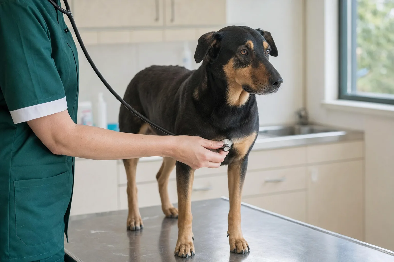
You are on the sofa, scratching his ribs, and your fingers find something soft under the skin, about the size of a walnut. It was not there last month. Or your nine-year-old Boxer has a reddish lump on his thigh that has grown noticeably since Easter. Or the year-old pup has a pink button on his eyelid. All three count as lumps, and you cannot identify any of them by eye or by touch. Neither can your vet. The answer comes from a fine needle and a microscope. Until then, your job is to measure, photograph, and know when a lump has stopped being a routine appointment.
When to call the vet today about a lump
Most lumps can wait for an ordinary appointment that week. A few cannot:
- it came up overnight and is hot, red and sore, and the dog is flat or feverish (usually an abscess after a bite, a sting or something lodged under the skin);
- it is bleeding, has burst, or has a scab that will not heal;
- it has visibly grown in two or three weeks, or changes size from one day to the next (some skin tumours swell up and go down again);
- you find several swellings at once, on both sides, under the jaw, in front of the shoulders, behind the knees, and the dog is eating less or tiring easily (those are lymph nodes);
- it is in the mouth, on the gum or under the tongue, especially if it is dark, or the dog is drooling and has bad breath;
- it is black or very dark on a toe, near a nail or on a lip;
- it sits on the mammary chain of an unspayed bitch and is getting bigger, or the gland is hot and hard;
- the belly has swollen over days rather than months;
- a whole leg has swollen up;
- alongside the lump, the dog has lost weight, is drinking more, is vomiting or is breathing hard.
On the phone, say where the lump is, when you first noticed it and how big it is. Compare it with something everyone knows: a pea, a cherry, a walnut. Say whether it has changed. If the practice takes photos, send one.
What to measure and note at home
You measure and keep track; the vet does the interpreting. You need a ruler or a cheap set of callipers, your phone and a notebook.
A lump map
Draw a rough dog on a sheet of paper, left side and right side. Every lump gets a cross, a date and a size in millimetres. At the next check you can see straight away what is new and what has grown. Take photos in daylight with the ruler next to the lump so the size shows.
Size, feel and the skin on top
Measure in two directions and write both numbers down. "Walnut-sized" means something different to everyone; 23 by 18 mm means the same to all of us. Measure again in a month unless your vet gives you a different interval.
Then note how it feels. Soft or firm? Does it slide freely under the skin like a marble in a pocket, or is it stuck to whatever is underneath? Is it in the skin (raising it, with or without hair on top) or under the skin (the skin moves over it)? Is it warm? Does the dog pull away when you touch it?
The skin over it can be normal, red, hairless, ulcerated, scabbed or black. Sometimes there is a pore in the middle that lets out something white and pasty.
Lymph nodes
Learn where they are while your dog is healthy: under the angle of the jaw, in front of the shoulder, behind the knee, in the groin. In a healthy dog they are small and hard to find. If they feel like grapes or bigger, write it down.
The monthly once-over
Once a month, when he is relaxed, run your hands over all of him. Head, neck, chest, belly, each leg down to between the toes, under the tail, and the mammary glands on a bitch. Finish by lifting his lips and looking in his mouth. It takes three minutes. Small lumps get found during careful stroking, rarely at bath time.
What else goes in the notebook
Breed matters: Boxers, Labradors, Golden Retrievers, Bulldogs and German Shepherds each have their own more common tumours. Note his age, whether he is neutered and when, whether he has had a vaccine or an injection in that exact spot recently, whether he has been in a scrap, and whether he keeps licking the area.
What it might be, going by looks
| What you see |
Commonly seen in |
What it might be |
| Soft, mobile, under the skin, slow-growing, not painful |
Adults and seniors, more often overweight dogs |
Lipoma (fat); still confirmed with a needle, because some tumours look the same |
| Smooth pink hairless button that came up quickly, on the head or a leg |
Dogs under 3 |
Histiocytoma; usually regresses on its own within a few months, once the needle has confirmed it |
| Small lump in the skin with a pore that lets out white paste, or a "wart" on an old dog |
Adults, seniors |
Cyst or benign tumour of the skin glands |
| Whitish cauliflower-like growth in the mouth or on the lips |
Puppies and young dogs |
Viral papilloma; usually clears up on its own |
| Hot, painful, red, came up overnight, dog subdued |
Any dog, after a fight or a sting |
Abscess |
| Any appearance; red, itchy, swells and shrinks |
Adults; Boxers, Bulldogs, Labradors |
Mast cell tumour, the most common malignant skin tumour in dogs and the best at disguising itself |
| Firm, fixed to the tissue below, slow-growing, on the body or a leg |
Adults, seniors, large breeds |
Soft tissue sarcoma |
| Along the mammary chain, one or several |
Unspayed bitches or bitches spayed late |
Mammary tumours; about half are malignant |
| Several enlarged lymph nodes at once, tired dog |
Middle-aged adults |
Lymphoma |
| Ear flap puffed up like a cushion, soft |
After a lot of head shaking |
Aural haematoma |
| Small grey-brown lump with "legs" |
Any dog, after long grass |
Tick; mistaken for a lump more often than you would think |
The table gives you the words to use with your vet. The needle gives the diagnosis: two lumps that look identical can be fat in one case and a tumour that needs surgery with wide margins in the other.
What the vet will do with a lump
- Ask questions. How long, how much it has grown, whether it changes, and whether anything is different about the dog: eating, drinking, energy. Your map and photos cut that conversation in half.
- Examine the whole dog, feel the lymph nodes and look for lumps you missed.
- Fine-needle aspirate. This is the step that decides things. A thin needle, like a vaccine needle, goes into the lump a few times; the cells are smeared on a slide and read under a microscope, in the practice or at a lab. No anaesthetic, a minute's work, and cheap next to anything else. Veterinary oncologists use a simple rule: any lump bigger than a centimetre that has been there for more than a month gets aspirated. Without the needle, "keeping an eye on it" means nothing.
- Biopsy, if the needle is not conclusive: a piece of tissue taken under sedation or anaesthetic and sent for histopathology. Within a few days you learn what it is and how aggressive it is (the grade). After surgery, the same lab tells you whether it all came out (the margins).
- Staging for a malignant tumour, meaning how far the disease has gone: blood tests, chest X-rays, an abdominal ultrasound, and an aspirate of the nearest lymph node. The results can change the whole plan.
- The plan. For most skin lumps, surgery is the treatment, with margins of healthy tissue chosen according to the tumour type. Ask for the removed tissue to go to histopathology even if it "looks benign". A lipoma is either left and measured or removed once it grows somewhere that interferes with movement, such as the armpit or chest. An abscess is opened and flushed under sedation. For aggressive or spread tumours, your vet will refer you to a veterinary oncologist, who will talk you through what chemotherapy and radiotherapy can offer, how much time they take and how the dog is likely to feel along the way. The decision is yours.
- Follow-up. After a malignant tumour, examinations and imaging at the intervals your vet sets. After a lipoma, your monthly measurement plus the annual check.
Before you leave the consulting room, make sure you know three things: what the needle showed, whether the tissue is going to histopathology, and when you are coming back.
What not to do at home
- Do not squeeze it, prick it or cut it. A squeezed cyst gets infected. Pressing on a mast cell tumour releases substances that make the area swell and can cause vomiting. An abscess lanced at home is left with the pocket still full.
- Do not reach for creams from the cupboard, disinfectants or "drawing" ointments, or internet recipes that claim to burn lumps off. A chemical burn hides exactly what needed to be seen and delays the diagnosis by weeks.
- Do not wait more than a few weeks to see if it goes away. A histiocytoma really does go on its own, but you only know it is a histiocytoma from the needle. After that you can wait calmly.
- Do not tell yourself it is "just another lipoma" because the last two were. Every new lump gets its own needle.
- Do not mistake a tick for a lump, or a lump for a tick. Look closely with your phone torch: a tick has legs and you can see where it is attached. Remove it with a tick hook, as close to the skin as you can, without squashing it.
- Do not accept a lump being "monitored" without an aspirate, whoever suggested it. Measuring monthly makes sense once you know what it is.
- Do not diagnose from photos online. A mast cell tumour can look like any row in that table, a lipoma included.
- Do not put off the neutering conversation with a young bitch. Spaying before the first season brings the risk of mammary tumours close to zero, and after the second season the protection has almost gone. Early neutering has its own downsides in some breeds, so pick the timing with your vet, for your dog.
- Do not drop the monthly once-over because "the vet saw him at his booster". A one-centimetre lump can come up in a few weeks.
If the lump has already been removed
Keep the histopathology report in his file with the date and the location. If it was malignant, ask which checks come next and how often, and put them in your diary there and then. Photograph the scar after a month and again at every check. Local recurrence tends to show up along the edge of the scar, and an old photo catches it sooner than your memory will.
Most lumps found on dogs are benign. The rule is the same for all of them: needle first, decisions after. We do our monthly once-over on nail-clipping day, which is the easiest way not to forget it. If you do not have a regular vet yet, our page on related services will help you choose a practice.
The information on this page is educational and does not replace an examination. For full details and a diagnosis, consult your dog's own veterinarian.